1Laboratorio de Anatomía Animal, Facultad de Medicina Veterinaria y Zootecnia, Universidad Central del Ecuador, Quito, Ecuador
2Facultad de Ingeniería Química, Universidad Central del Ecuador, Quito, Ecuador
3Grupo de Investigación en Alimentos, Compuestos Orgánicos, Materiales, Microbiología Aplicada y Energía (ACMME), Universidad Central del Ecuador, Quito, Ecuador
4Laboratory of Plastination and Anatomical Techniques, Universidad de La Frontera, Temuco, Chile
Teaching morpho-functional sciences requires anatomical specimens, but traditional formaldehyde preservation poses health risks. Plastination provides safe, durable samples but is limited by cost and regulatory restrictions on solvents and polymers. This pilot study evaluated ethanol, thinner, and platinum silicone as alternatives for producing plastinated specimens in Ecuador. We applied Von Hagens’ plastination protocol to chicken organs (heart, gizzard, and proventriculus), adapting the dehydration–degreasing and forced impregnation phases. Organs were fixed in 10% formaldehyde for 15 days. Alternative solvents (ethanol, acetone, thinner) were tested at -20 °C; ethanol and acetone were selected for minimal volumetric shrinkage. Impregnation compared Biodur® (S10:S3) with platinum silicone, followed by curing with S6 Biodur® or manual catalyst application. We recorded volumetric changes after dehydration and at process completion. Ethanol and acetone yielded lower shrinkage in hearts (12%) and gizzards (1–3%) versus thinner (43%). Qualitatively, both organs processed with Biodur® silicones (BS) and those treated with platinum silicone (PS) were dry, odorless, retained their overall anatomical shape, and could be handled without structural damage. However, the present study did not include a standardized scoring system for specimen quality assessment, which represents a limitation and should be addressed in future studies. In addition, the Bayesian statistical analyses of volumetric shrinkage did not provide sufficient evidence of differences between the two polymers. Consequently, while these results may suggest a possible trend toward the technical feasibility of platinum silicone as an alternative, further in-depth studies with larger sample sizes are required. Nevertheless, the findings of this pilot study support the potential feasibility of an alternative dehydration protocol using ethanol.
ethanol; food-grade silicone; impregnation; volumetric shrinkage
María Revelo-Cueva, Laboratorio de Anatomía Animal de la Universidad Central del Ecuador, Jerónimo Leyton S/N y Gatto Sobral, Quito, Ecuador
e-mail: mcrevelo@uce.edu.ec





In medical education, the use of specimens such as whole bodies, organs, joint preparations, and isolated sections is required (Beltrán, 2010; Muñetón, 2012). To use them efficiently in anatomy laboratories, preservation techniques are employed (Muñetón, 2012). Traditional anatomical preservation methods rely on formaldehyde, which poses a health risk not only because it irritates the ocular and nasal mucosa but also because it is potentially carcinogenic (Espinosa et al., 2006; Muñetón, 2012; Popp et al., 2018).
Plastination is a preservation technique that produces anatomical specimens free of formaldehyde (thus safer), with extended durability and high educational value (Fonseca, 2017; Prieto et al., 2019; López et al., 2021; Ottone, 2021; Ottone, 2023). These advantages have contributed to its continued use over time (Muñetón, 2012; Popp et al., 2018). Nevertheless, the trademark polymers employed during the forced impregnation stage of von Hagens’ standard technique are difficult to import (Muñetón, 2012; Fonseca, 2017). Likewise, acquiring the organic solvents used in the dehydration–defatting process is challenging, as they are classified as controlled substances because of their potential use in the illegal manufacture of narcotics or because they require complex handling in laboratory settings (Fonseca, 2017; Acevedo et al., 2018).
In various studies, solvents such as isopropyl alcohol, 1,1,1-trichloroethane, and ethyl alcohol have been used as alternatives for organ dehydration (Singh et al., 2015; Fonseca, 2017; Peralta, 2017; Acevedo et al., 2018). Nonetheless, each solvent presents specific considerations for its use. For instance, isopropyl alcohol, like acetone, is classified as a controlled substance (Acevedo et al., 2018). Similarly, 1,1,1-trichloroethane, due to its similarity to chloroform, has been prohibited for importation into Ecuador since 2015 (Comité de Comercio Exterior del Ecuador, 2019). For these reasons, ethyl alcohol (ethanol) remains a safe and viable alternative.
For organ defatting, acetone or methylene chloride is conventionally used (Ottone, 2021; Ottone, 2023). Acetone is restricted due to its classification as a controlled substance (Acevedo et al., 2018). Meanwhile, methylene chloride is potentially hazardous and poses significant risks during laboratory handling. Consequently, alternative degreasing agents must be considered. Thinner is commonly used as a degreasing agent and is typically used to dilute enamels or lacquers. Although no literature documents the use of thinner in plastination techniques, its availability in the Ecuadorian market and relatively low cost make it a feasible option.
Regarding the replacement of silicones in plastination, several researchers have explored alternative materials. In Brazil, P10 silicone (Polisil Inc.) was used as an alternative to Biodur® S10 silicone (Monteiro et al., 2016). Similarly, in Argentina, the standard-protocol silicone was replaced with a household silicone containing ethyl triacetoxysilane (Fastix®, Akapol) (Audisio et al., 2021). However, some studies have also replaced commercial silicones with more economical materials, such as an emulsion obtained by dissolving plastic Petri dishes in xylene (Singh, 2015). These studies may give the impression that replacing silicones is relatively “simple”; nevertheless, the complete chemical characterization of the silicones used in von Hagens’ standard protocol was reported only in 2004 (Chaynes & Mingotaud, 2004). This knowledge makes it possible to develop protocols using commercially available silicones in each country. Conversely, modifying both the dehydrating agent and the silicone type requires optimizing new protocols, which may be associated with increased organ shrinkage (Monteiro et al., 2022; Delpupo et al., 2024).
The Animal Anatomy Laboratory at the Universidad Central del Ecuador (UCE) houses Ecuador's only plastination laboratory, and the anatomical specimens processed there are used not only as study material but also in the creation of interactive atlases (http://atlascanino.uce.edu.ec/organos/). To continue producing anatomical models in UCE’s plastination area, it is necessary to identify alternative organic solvents for the dehydration–defatting phase and to test polymers that can be sourced from the national market. Therefore, this pilot study aimed to use ethyl alcohol and thinner as alternatives for the dehydration–defatting process, along with a food-grade or platinum silicone (PS) available in the Ecuadorian market.
The protocol described by von Hagens (von Hagens, 1979; von Hagens et al., 1987) was employed for the preservation of organs, including the heart, gizzard and proventriculus, from domestic chickens (Gallus gallus domesticus). The plastination technique consists of four phases: (1) fixation, (2) dehydration–defatting, (3) forced impregnation, and (4) curing. However, in the present study, modifications were introduced in the dehydration–defatting and forced impregnation phases. Specifically, alternative solvents—ethyl alcohol and thinner—were tested for the dehydration–defatting phase, whereas platinum-based silicone (PS) was used for the forced impregnation phase.
In this phase, hearts, gizzards and proventriculi from domestic chickens were used, obtained directly from certified supermarkets authorized by the Metropolitan District of Quito. All organs originated from clinically healthy domestic chickens processed under official sanitary control. Approval from the Animal Research Ethics Committee (CEIA) was unnecessary for this study, because ethical clearance is only granted for research involving live vertebrate animals and the study was conducted on organs intended for consumption. Furthermore, efforts were made to select specimens with similar weight within each experimental repetition to promote homogeneity among samples. Prior to inclusion, all organs were macroscopically evaluated to ensure the absence of visible pathological alterations. Despite these measures, inherent biological variability in tissue composition and structure cannot be eliminated.
The organs were washed and dissected. Each heart, gizzard and proventriculus was then weighed using a Truper digital scale (range: 1 g to 5 kg) and identified with an alphanumeric code. Additionally, width, length, and thickness were recorded to determine the volume of each organ. Subsequently, the organs were immersed in a 10% formaldehyde fixing solution. The glass containers used for fixation were equipped with airtight lids, and a layer of cotton was placed at the bottom of each container beforehand to prevent direct contact between the organs and the container base. The organs were fixed for 15 days.
This phase was carried out in two sub-stages: 2.1 Experimentation using alternative solvents.
2.2 Dehydration–defatting of domestic chicken organs following the protocol established during the experimental substage.
2.1. Experimentation using alternative solvents
For the experimental phase, two pilot tests were conducted. Two hearts and two gizzards were used for each proposed test:
2.2. Dehydration–defatting of domestic chicken organs using solvents selected during the experimental substage
Based on the lower volumetric shrinkage percentages obtained in the first dehydration–defatting test, ethyl alcohol and acetone were selected. A total of 18 organs (6 hearts, 6 gizzards, and 6 proventriculi) from domestic chicken fixed in 10% formalin for 12 days were used. Dehydration was performed with ethyl alcohol in ascending concentrations of 70%, 96%, and 99% at –20 °C, with replacements every 48 hours. Between each alcohol change, its concentration was measured, and once it remained at 99% for two consecutive measurements, the organs were transferred to acetone for 24 hours at room temperature.
At this stage, the organs were divided into two experimental groups. The control group (3 hearts, 3 proventriculi, and 3 gizzards) was processed using a mixture of Biodur® silicones (BS) S10 and S3 in a 100:1 ratio. In contrast, the experimental group used platinum silicone (PS) or food-grade silicone (DSH 20, MOLMA), translucent, with a viscosity of 7000 cPs (centipoise), consisting of two components (A and B) that must be mixed in equal parts (1:1) when used for mold preparation. Considering that component B acts as the silicone catalyst, a 10:1 ratio was used instead of the manufacturer’s recommended preparation. This modification of the silicone-to-catalyst ratio constituted an experimental adjustment intended to extend the working time during impregnation. The 10:1 ratio was selected based on a preliminary trial, in which lower catalyst concentration was found to delay polymerization, thereby facilitating better handling during impregnation.
Air was removed from both silicone mixtures using vacuum pressure with a 1.5 m³/h vacuum pump (Slaykov u Brna, model TYP JMC63-4S B5). Subsequently, the dehydrated organs were fully immersed in each mixture separately. Each mixture was contained in individual 2 quarts of stainless-steel bain-marie pots, which were placed inside a 35 L Heidelberg Plastination Kettle (Biodur®).
The impregnation process began at a pressure of 80 mmHg at −20 °C, using an adapted freezer equipped with copper tubing for the vacuum system. As bubble formation ceased, the pressure was gradually reduced until reaching 1 mmHg; pressure was monitored and controlled using a digital manometer (Greisinger, model GDH 12 AN) and vacuum adjustment valves. The impregnation process lasted two weeks, and in the case of platinum silicone (PS), it was necessary to manually remove residual silicone adhering to the organs using paper towels.
The curing process for organs impregnated with Biodur® S10:S3 was performed using Biodur® S6. For this purpose, the organs were placed in a Biodur® curing chamber, model HH13A1.0, which was operated in four-hour cycles. In contrast, the curing process for organs impregnated with the platinum-based food-grade silicone was carried out by daily brushing component B onto the surface of each organ. After one week of curing, all organs were stored in plastic containers.
Given the limited sample size (n = 3 per group), traditional frequentist tests did not meet statistical assumptions. Instead, a Bayesian estimation approach (BEST) was applied. This method is suitable for exploratory analysis in small samples; however, it does not overcome the limitations associated with low statistical power. Thus, the results were interpreted with caution. Given the analysis involved a basic Bayesian statistical comparison between two small groups, Microsoft Excel was used.
A comparison between two independent groups was performed: organs impregnated with platinum silicone (PS) and organs processed with Biodur® silicones (BS). For each group, a normal likelihood with unknown mean (μ) and variance (σ2) was assumed, using a non‑informative Jeffreys prior. The posterior model applied was the conjugate model. Consequently, the conditional distributions given the data (y) followed known analytical expressions. The conditional posterior variance adopted an Inverse Chi-squared distribution (Inverse–χ²) with degrees of freedom (ν) equal to n − 1 and a scale parameter equivalent to the sample variance (s²):
σ² | y ~ Inverse–χ² (ν = n − 1, τ² = s²)
In turn, the conditional posterior mean followed a normal distribution with mean equal to the observed sample mean and variance proportional to σ²/n.The joint posterior distribution was generated through extensive simulations (≥ 200,000 iterations), using a fixed random seed to ensure reproducibility. From these simulations, the posterior means for each group (μ_PS and μ_BS) were estimated. The group comparison was then conducted using the posterior difference of means. In addition, the standardized effect size was calculated using Hedges’ g, including the small-sample correction to avoid bias. For this calculation, the pooled standard deviation (SD_pooled) was used: g = (μ_PS − μ_BS) / SD_pooled
The hearts of Gallus gallus domesticus dehydrated with ascending concentrations of ethyl alcohol and degreased with acetone exhibited a mean volumetric shrinkage of approximately 12% and an average weight reduction of 3 g. The gizzards showed shrinkage between 1% and 3%, accompanied by a weight decrease of 8 to 10 g (Table I).
| No. | Organ | Code | Initial weight (g) | Weight after dehydration (g) | Weight reduction (%) |
Initial volume (cm3) |
Post-dehydration volume(cm3) | Volumetric shrinkage (%) |
| 1 | Heart | 1H-ea | 15.00 | 12.00 | 20.00 | 5.89 | 5.18 | 12.11 |
| 2 | Heart | 2H-ea | 16.00 | 13.00 | 18.75 | 8.40 | 7.39 | 12.00 |
| Mean | 19.38 | Mean | 12.05 | |||||
| 1 | Gizzard | 1G-ea | 38.00 | 28.00 | 26.32 | 7.75 | 7.64 | 1.35 |
| 2 | Gizzard | 2G-ea | 31.00 | 23.00 | 25.81 | 6.70 | 6.49 | 3.13 |
| Mean | 26.06 | Mean | 2.24 | |||||
H: heart; G: gizzard; ea: ethanol plus acetone
b. Dehydration with ethyl alcohol and degreasing with thinner:
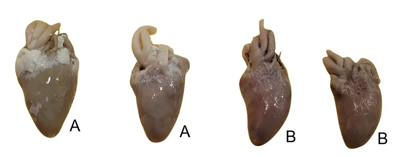
Figure 1. Hearts of domestic chickens dehydrated and degreased using alternative solvents.
Pictures (A) show hearts dehydrated with ethanol and degreased with acetone. Pictures (B) display hearts dehydrated with ethanol and degreased with thinner, where color changes, degree of visual degreasing, and reduced size of anatomical specimens can be observed.
Dehydration performed with ethyl alcohol combined with degreasing using thinner (LACA Nitro, Pintuco) resulted in greater shrinkage in the hearts, with an average of 43.46%, while the gizzards showed an average shrinkage of 8.85% (Table II), suggesting a possible trend toward greater shrinkage in the hearts under this treatment. However, it was noted that the hearts treated with thinner (LACA Nitro, Pintuco) exhibited more visibly pronounced degreasing compared with those immersed only in ethyl alcohol (Fig. 1). Nevertheless, the composition of commercial thinner products may vary among manufacturers and regions; therefore, this should be considered a limitation, as such variability may affect the reproducibility of the procedure and the resulting tissue changes when different commercial formulations are employed.
| No. | Organ | Code | Initial weight (g) |
Weight after dehydration (g) | Weight reduction (%) | Initial volume (cm3) |
Post-Dehydration volume (cm3) |
Volumetric shrinkage (%) |
| 1 | Heart | 1H-et | 12.00 | 9.00 | 25.00 | 6.48 | 3.63 | 43.98 |
| 2 | Heart | 2H-et | 11.00 | 8.00 | 27.27 | 6.45 | 3.68 | 42.95 |
| Mean | 26.14 | Mean | 43.46 | |||||
| 1 | Gizzard | 1G-et | 26.00 | 9.00 | 65.38 | 5.55 | 5.13 | 7.55 |
| 2 | Gizzard | 2G-et | 25.00 | 8.00 | 68.00 | 7.23 | 6.49 | 10.14 |
| Mean | 66.69 | Mean | 8.85 | |||||
H: heart; G: gizzard; et: ethanol plus thinner
Considering the trend suggesting greater shrinkage caused by the ethanol–thinner dehydration–degreasing procedure in the hearts, ethyl alcohol was selected for the dehydration step, while acetone was utilized for the degreasing process.
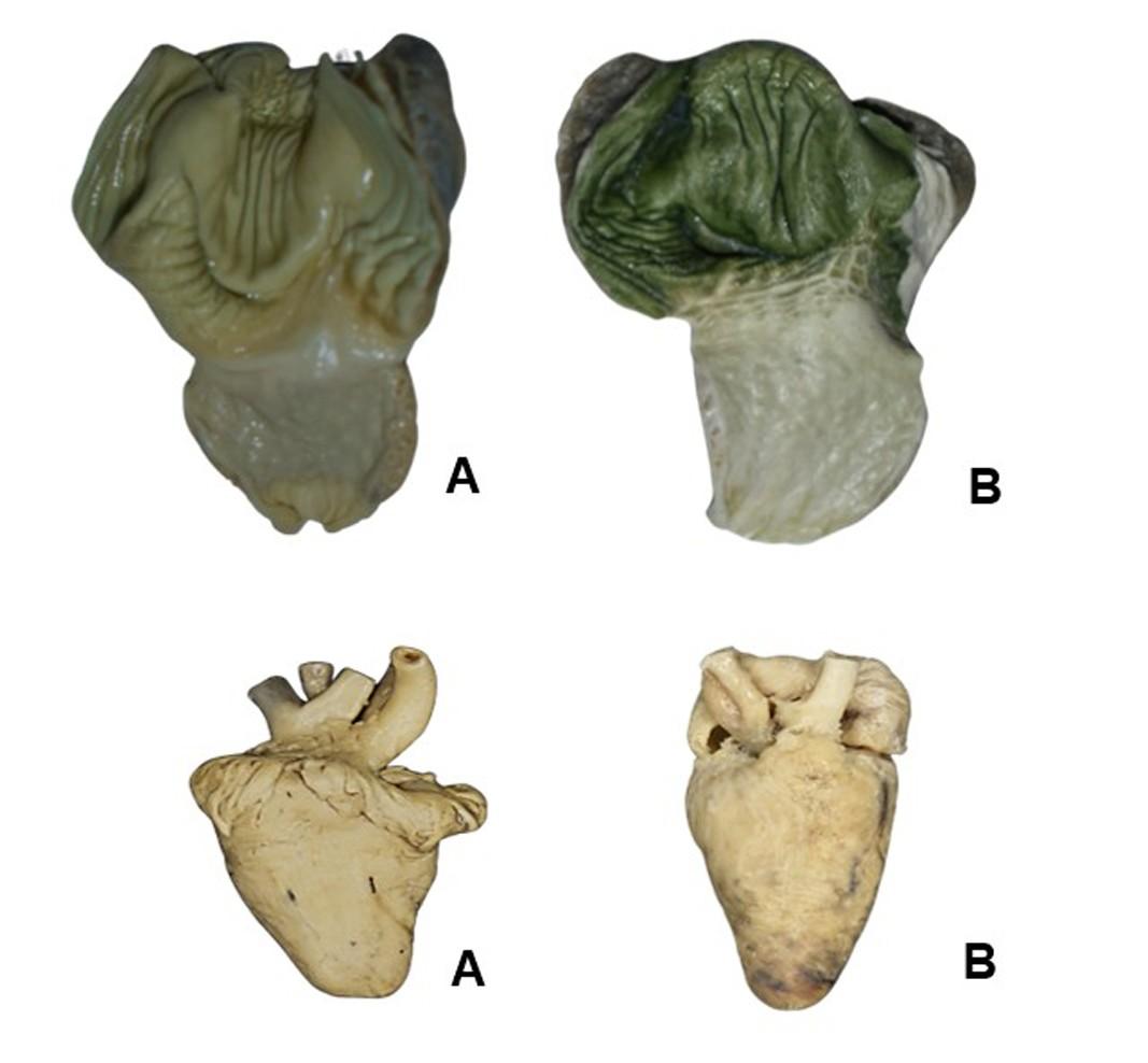
Figure 2. Heart, gizzard, and proventriculi process using both Biodur® silicone (A) and the tested platinum silicone (B)
Three hearts, three proventriculi, and three gizzards were successfully plastinated with each type of silicone used. From a visual standpoint, both the organs processed with Biodur® silicones (BS) and those treated with platinum silicone (PS) were dry, odorless, and exhibited a glossy appearance (Fig. 2).
Furthermore, average volumetric shrinkage for hearts was 30.41% with BS and 31.89% with PS (Table III). When the Bayesian statistical comparison was performed between groups, it suggested small differences accompanied by substantial uncertainty. The posterior means for BS and PS (μ_BS and μ_PS, respectively) showed extensive overlap, and the posterior difference (Δ) had a mean of 1.38 p.p. (Table IV). The posterior probability that PS exhibited greater shrinkage than BS was 56.65%, a value close to 0.50 that does not support a clear advantage of either treatment (Table IV). Consistently, the corrected Hedges’ g was 0.11, which is expected in a pilot study with a very small sample size such as the one conducted here (n=3). Overall, the results did not provide clear evidence of differences in volumetric shrinkage of hearts between PS and BS.
| No. | Code | Initial volume (cm3) | Volume after dehydration (cm3) | Volumetric shrinkage (%) |
| 1 | 1H-BS | 3.82 | 2.65 | 30.73 |
| 2 | 2H-BS | 3.36 | 2.52 | 25.00 |
| 3 | 3H-BS | 3.41 | 2.20 | 35.48 |
| Mean | 30.41 | |||
| 1 | 1H-PS | 3.50 | 2.80 | 19.91 |
| 2 | 2H-PS | 3.38 | 2.11 | 37.53 |
| 3 | 3H-PS | 3.68 | 2.27 | 38.22 |
| Mean | 31.89 |
Regarding gizzards, the average volumetric shrinkage was calculated (Table V), and the Bayesian statistical comparison showed that the posterior means for BS and PS were 26.43% and 31.05%, respectively, with substantial overlap of their credible intervals (Table VI). The posterior difference (Δ) had a mean of 4.63 p.p. (Table VI).
| No. | Code | Initial volume (cm3) |
Volume after dehydration (cm3) |
| 1G-PS | 7.75 | 4.50 | 35.82 |
| 2G-PS | 7.02 | 4.50 | 31.34 |
| 3G-PS | 4.82 | 3.56 | 26.09 |
| Mean | 31.08 | ||
| 1G-BS | 7.44 | 5.65 | 23.94 |
| 2G-BS | 7.02 | 4.82 | 31.34 |
| 3G-BS | 4.82 | 3.67 | 23.91 |
| Mean | 26.40 |
The posterior probability of superiority was 0.8, suggesting a possible trend toward greater shrinkage in gizzards impregnated with PS compared with those processed with BS; however, the substantial uncertainty and wide credible intervals preclude definite conclusions. The corrected Hedges’ g was 0.60, with a very wide 95% credible interval (−0.78 to 2.01). Taken together, and considering the breadth of the intervals, greater shrinkage in gizzards plastinated with PS compared with those treated with BS remains only a possibility that should be verified in studies with larger sample sizes.
| No. | Code | Initial volume (cm3) |
Volume after dehydration (cm3) |
| 1P-PS | 3.98 | 3.67 | 7.89 |
| 2P-PS | 3.98 | 3.67 | 7.89 |
| 3P-PS | 1.47 | 1.26 | 14.29 |
| Mean | 10.03 | ||
| 1P-BS | 3.56 | 3.14 | 11.76 |
| 2P-BS | 3.35 | 2.93 | 12.50 |
| 3P-BS | 1.47 | 1.05 | 28.57 |
| Mean | 17.61 |
| Measure | Group / Definition | Posterior Mean | CI 95% (lower; upper) |
Notes |
| Volumetric shrinkage (%) | BS | 26.43 | 15.78; 37.01 | |
| Volumetric shrinkage (%) | PS | 31.05 | 18.98; 43.18 | |
| Difference (p.p.) | Δ = μ_PS − μ_BS | 4.63 | −12.82; 22.02 | |
| Probabilities (%) | Pr (Δ > 0) | 80.17 | — | |
| Pr (Δ < 0) | 19.83 | — | ||
| Effect size | Hedges g (uncorrected) | 0.75 | −0.98; 2.51 | |
| Hedges g (corrected) | 0.60 | −0.78; 2.01 | J = 0.80 |
Δ: posterior difference between groups (Δ); credible interval (CI); percentage points (p.p.); small‑sample correction factor in Hedges’ g (J)
Finally, on average, the volumetric shrinkage of proventriculi impregnated with PS was 10.03%, whereas it was 17.61% for those processed with BS (Table VII). Furthermore, the posterior mean shrinkage for the proventriculi was 17.68% for BS and 10.00% for PS. The posterior difference (Δ) was −7.68 p.p. (Table VIII).
| No. | Code | Initial volume (cm3) | Volume after dehydration (cm3) |
| 1P-PS | 3.98 | 3.67 | 7.89 |
| 2P-PS | 3.98 | 3.67 | 7.89 |
| 3P-PS | 1.47 | 1.26 | 14.29 |
| Mean | 10.03 | ||
| 1P-BS | 3.56 | 3.14 | 11.76 |
| 2P-BS | 3.35 | 2.93 | 12.50 |
| 3P-BS | 1.47 | 1.05 | 28.57 |
| Mean | 17.61 |
The posterior probability of superiority of PS over BS was 18.06% (Pr (Δ < 0) = 81.94%), and the corrected Hedges’ g was −0.66 (Table VIII). This indicates a possible tendency toward lower volumetric shrinkage in proventriculi impregnated with PS compared with those processed with BS; however, the wide credible intervals prevent this from being interpreted as a definitive finding.
| Measure | Group / Definition | Posterior Mean | CI 95% (lower; upper) |
Notes |
| Volumetric shrinkage (%) | BS | 17.68 | -5.95; 41.17 | |
| Volumetric shrinkage (%) | PS | 10.00 | 0.84; 19.20 | |
| Difference (p.p.) | Δ = μ_PS − μ_BS | -7.68 | -33.93; 18.89 | |
| Probabilities (%) | Pr (Δ > 0) | 18.06 | — | |
| Pr (Δ < 0) | 81.94 | — | ||
| Effect size | Hedges g (uncorrected) | -0.83 | -2.62; 0.93 | |
| Hedges g (corrected) | -0.66 | 2.09; 0.74 | J = 0.80 |
Δ: posterior difference between groups (Δ); credible interval (CI); percentage points (p.p.); small‑sample correction factor in Hedges’ g (J)
The present pilot study suggests the technical feasibility of implementing an alternative plastination protocol for organs of domestic chickens (Gallus gallus domesticus) using solvents commercially available in Ecuador. Specifically, replacing acetone with ethyl alcohol as the dehydrating agent in the plastination protocol may represent a viable, albeit not optimal, alternative in countries with limited access to controlled solvents (Srisuwatanasagul et al., 2010; Singh et al., 2015; Acevedo et al., 2018). The results showed that dehydration with ascending concentrations of ethyl alcohol (70%, 96%, 99%) at –20 °C, followed by degreasing with acetone, produced acceptable volumetric shrinkage in gizzards (1–3%), although higher in hearts (12%). In contrast, previous studies have reported greater tissue shrinkage with ethanol compared to pure acetone (Srisuwatanasagul et al., 2010; Singh et al., 2015).
The incorporation of thinner as a degreasing agent, for which there is limited precedent in literature, was associated with greater shrinkage in hearts (43.46%) and lower shrinkage in gizzards (8.85%). This difference may be attributable to distinctive histological composition: the myocardium consists of striated muscle tissue with a complex fibrillar structure, whereas the gizzard contains dense smooth muscle and a cuticular layer that confers greater resistance to shrinkage (Troup & Stein, 1995; Monteiro et al., 2022). However, the use of commercial thinner should be interpreted with caution, since its chemical composition may differ depending on the manufacturer, formulation, and region of availability. Such variability could influence the degree of degreasing, tissue shrinkage, and overall reproducibility of the procedure.
Concerning the use of platinum silicone (PS) as a possible alternative, the organs obtained showed generally comparable characteristics in terms of odor absence and bright visual appearance. Nevertheless, the Bayesian statistical analysis suggested greater shrinkage in hearts and gizzards processed with PS. Monteiro et al. (2022) reported that polymer viscosity directly influences tissue shrinkage, with lower-viscosity silicones producing less contraction compared to S10 (Biodur®). Given that the PS used in this pilot study has a viscosity of 7000 cPs, this property may be related to the apparently greater shrinkage observed. In contrast, proventriculi processed with Biodur® silicones exhibited apparently greater volumetric shrinkage according to the Bayesian analysis. However, it is important to consider that, although all organs were sourced from certified suppliers, selected to have similar weights, and macroscopically screened to exclude visible pathological alterations, some degree of biological variability is unavoidable. In addition, because this was a pilot study with a small sample size, these findings should be interpreted as preliminary evidence regarding the feasibility of PS.
Furthermore, volumetric shrinkage occurs predominantly during the forced impregnation phase when the intermediary solvent is replaced by the polymer (Monteiro et al., 2022; Delpupo et al., 2024). This phenomenon was demonstrated by comparing shrinkage percentages after dehydration and after impregnation–curing. The results suggest that alternative dehydrating agents and silicones may increase organ shrinkage, particularly in hearts and gizzards, where values exceeded 30%. Such shrinkage may potentially alter anatomical proportions, an important consideration given the increasing reliance on plastinated avian models, such as chicken, for teaching gross anatomy, where maintaining structural fidelity is highly beneficial for student learning (Troup & Stein, 1995; Muñetón & Ortiz, 2012; Ottone et al., 2020; López et al., 2021; Oliver & Adds, 2023; Delpupo et al., 2024). Therefore, one methodological limitation acknowledged in this study is the absence of microscopic measurements. Histological characterization not only evaluates microscopic preservation after plastination but also determines whether an alternative protocol preserves the fine morphological integrity required for advanced educational or research applications (Popp et al., 2018; Monteiro et al., 2022; Monteiro et al., 2025).
The use of alternative polymers for plastination remains a relevant challenge, particularly considering the specific chemical composition and material properties of the reagents originally developed for this technique. Chaynes and Mingotaud (2004) reported that Biodur® S10 corresponds to polydimethylsiloxane (PDMS), Biodur® S3 to dibutyltin dilaurate, and Biodur® S6 to tetraethyl orthosilicate. Although this chemical characterization could support the development of alternative protocols, substitution has not been straightforward. Several researchers have tested substitutes that differ from the specialized chemicals designed for plastination, obtaining acceptable results depending on local reagent availability (Singh, 2015; Monteiro et al., 2016; Audisio et al., 2021). Henry et al. (2019) emphasized that Biodur® silicones were specifically developed for optimal plastination, explaining their widespread use. Accordingly, although organs processed with PS showed apparently normal physical characteristics, the observed shrinkage could potentially affect anatomical accuracy. In this regard, PS is an addition-cured elastomer with a platinum catalyst, whereas PDMS (Biodur® S10) is a linear fluid polymer that reticulates through the action of S3 and S6. Thus, despite both being siloxane derivatives, they differ considerably in their material properties (a silicone oil versus a silicone rubber) (Chaynes & Mingotaud, 2004; Pulido et al., 2011). Therefore, another limitation of the present pilot study is related to the type of polymer employed as an alternative.
Finally, there are also technical limitations associated with PS. The manual removal of silicone after impregnation suggests differences in adhesion and tissue penetration (Monteiro et al., 2016; Weiglein & Henry, 1993). Furthermore, the Biodur® protocol employs standardized curing procedures using S6 gas in sealed chambers, whereas PS requires daily manual catalyst application (Troup & Stein, 1995; Fonseca, 2017; López et al., 2021; Monteiro et al., 2025). Additionally, the high viscosity of PS limits its reuse, increasing both costs and operational difficulties.
This pilot study demonstrates the technical feasibility of an alternative plastination protocol, using ethyl alcohol and platinum silicone (PS) as accessible substitutes for conventional reagents. The use of PS produced plastinated specimens with satisfactory quality, stability, and handling characteristics, suggesting that PS may represent a viable option for plastination in settings with limited access to specialized polymers traditionally required for this technique; however, further studies are needed to confirm its performance and broader applicability. In addition, the results obtained using ethyl alcohol are consistent with previous studies, supporting its feasibility as a dehydration agent within the plastination process. Its availability, lower regulatory restrictions, and reduced cost may help overcome logistical and economic barriers in countries such as Ecuador, facilitating broader implementation of plastination protocols without significantly compromising the quality of the resulting specimens.
Crucially, this approach contributes to democratizing access to plastination by enabling resource-limited laboratories to produce durable and safe anatomical specimens for teaching and research purposes. This is particularly relevant in regions where access to controlled solvents and commercial plastination reagents is restricted. Although the small sample size limits the generalizability of the conclusions, the present findings provide a robust foundation for future studies involving larger samples and further methodological refinements. Overall, the proposed protocol represents a promising step toward the development of more accessible, adaptable, and sustainable plastination techniques.
Acknowledgments
Supported by the Research Directorate of the Universidad Central del Ecuador through the senior research project coded DI-CONV-2023-003.
Acevedo LM, Rojas MA, Velásquez JM. 2018: Técnica de plastinación de la Universidad de Antioquía: una adaptación del modelo estándar alemán [Plastination technique Antioquia University: an adaptation of the German standard method]. Iatreia Julio-septiembre 31(3): 228-239. doi:10.17533/udea.iatreia.v31n3a01.
Audisio S, Vaquero P, Ocampo L, Mondino M, Guinta J. 2021: Plastinación de una laucha de laboratorio con silicona de uso doméstico: comunicación de un ensayo [Plastination of a laboratory mouse with a domestic use silicone: case report]. Revista Académica de Investigación, Docencia y Extensión de las Ciencias Veterinarias 2(2): 20-25. https://ojs.unlpam.edu.ar/ojs/index.php/Vetec/issue/view/463.
Beltrán JA. 2010: La plastificación en la Universidad Nacional de Colombia-primera parte [Plastination at the National University of Colombia – Part One]. Morfolia 2(1): 3-17. https://revistas.unal.edu.co/index.php/morfolia/article/view/98833
Comité Comercio Exterior del Ecuador-COMEX. 2019: Resolución 031-2019. URL: https://www.produccion.gob.ec/wp-content/uploads/2019/12/Resoluci%C3%B3n-COMEX-031-2019.pdf [november, 2025].
Chaynes P, Mingotaud AF. 2004: Analysis of commercial plastination agents. Surg Radiol Anat 26(3): 235-238. doi: 10.1007/s00276-003-0216-9.
Delpupo F, Cassiano L, Monteiro Y, Júnior M, Soares K, Bittencourt A. 2024: Low viscosity silicone with less shrinkage for brain slices. Morphologie 108(360): 100726. doi: 10.1016/j.morpho.2023.100726.
Espinosa MT, Rojas MP, Bernal ML, Araque A, Vélez M, López JM. 2006: Manual of IARC Group 1 and 2A Carcinogenic Agents of Occupational Significance in Colombia. Manual de agentes carcinógenos de los grupos 1 y 2A de la IARC, de interés ocupacional para Colombia. URL: https://www.minsalud.gov.co/sites/rid/Lists/BibliotecaDigital/RIDE/IA/INCA/Manual-agentes-carcinogenos-2006.pdf [march, 2025].
Fonseca J. 2017: La técnica de plastinación, sus fundamentos y alternativas de menor costo [Plastination technique, fundamentals and cheaper alternatives]. Gaceta de Ciencias Veterinarias 22(2): 43-47. https://revistas.uclave.org/index.php/gcv/article/view/321
Henry RW, von Hagens G, Seamans G. 2019: Cold temperature/Biodur®/S10/von Hagens'—Silicone plastination. Anat Histol Embryol 48(6): 532-538. doi: 10.1111/ahe.12472.
López O, Gil F, Latorre R. 2021: Plastinated Organs as an Innovation Resource for STEAM Education. Órganos plastinados como recurso de innovación para la educación STEAM. Revista Internacional para la calidad educativa 2(1): 86-99. https://revistaeduca.org/educa/article/view/8
Monteiro Y, Laissa J, Bittencourt A, Siqueira B, Baptista C, Bittencourt A. 2018: Influence of the temperature on the viscosity of different types of silicone. J Plast 30(1): 17. doi: 10.56507/HETT9088.
Monteiro Y, Silva M, Bittencourt A, Bittencourt A. 2022: Plastination with low viscosity silicone: strategy for less tissue shrinkage. Braz J Med Biol Res 55: e11962. doi: 10.1590/1414-431X2022e11962.
Monteiro YF, Campos LESB, Patrício EA, Bittencourt AS, Bittencourt AS. 2025: A new epoxy for plastination: feasibility and applicability in Brazil. Braz J Med Biol Res 58: e13638. doi:10.1590/1414-431X2025e14839
Muñetón A, Ortiz A. 2012: Plastination: A complementary tool for the development of the teaching-learning process of anatomy. Rev Med Vet 23: 111-117. http://www.scielo.org.co/scielo.php?script=sci_arttext&pid=S0122-93542012000100011&lng=en&tlng=
Olivier G., Adds P. 2023: Chicken plastination: Its role in teaching avian anatomy. J Plast 35(1): 9. doi: 10.56507/GXLG9124
Ottone NE, Guerrero M, Alarcón E, Navarro P. 2020. Statistical analysis of shrinkage levels of human brain slices preserved by sheet plastination technique with polyester resin. Int J Morphol 38(1):13-16. doi: 10.4067/S0717-95022020000100013
Ottone NE. 2021: Protocolo unificado de plastinación con silicona en frío y a temperatura ambiente [Unified plastination protocol with silicone at cold and room temperature]. Int J Morphol 39(2): 630-634. doi:10.4067/S0717-95022021000200630
Ottone NE. 2023: Advances in Plastination Techniques. Cham; Springer Nature. doi: 10.1007/978-3-031-45701-2
Peralta E, Beltrán J, Luque R, Quijano Y. 2017: La plastinación como técnica de preservación de material biológico para docencia e investigación en anatomía [Plastination as a technique for preserving biological specimens for teaching and research in anatomy]. Morfolia 9(1): 55-62. https://repository.urosario.edu.co/handle/10336/28386.
Popp AI, Basso AP, Lodovichi MV, Sidorkewiej NS. 2018: Conservation of body sections and organs of the narrownose smooth-hound, Mustelus schmitti (Pisces, Chondrichthyes), by silicone injection at room temperature to be used in comparative anatomy learning. Int J Morphol 36(2): 413-418. doi:10.4067/S0717-95022018000200413.
Prieto R, Vargas CA, Veuthey C, Aja-Guardiola S, Ottone N. 2019: Fundamental concepts of the modified room temperature plastination protocol with silicone, with subsequent pigmentation, and its application for the conservation of human placenta. Int J Morphol 37(1): 369-374. doi:10.4067/S0717-95022019000100369.
Pulido JE, Salazar R, Gómez O, Sarmiento LR, Meza J, Navarro O. 2011: Preparación y evaluación de compatibilizantes para mezclas de poliesterno/silicona y caucho natural/silicona [Preparation and evaluation of compatibilizing agents for polystyrene/silicone and natural rubber/silicone blends]. Revista ION 17(1).
https://revistas.uis.edu.co/index.php/revistaion/article/view/1303.
Singh N, Chaudhary A, Nair S, Kumar S. 2015: Non-perishable museum specimens: redefined plastination technique. J Plast 27(2): 1-9. doi:10.56507/BRET3411.
Srisuwatanasagul K, Srisuwatanasagul S, Adirekthaworn A, Darawiroj D. 2010: Comparative study between using acetone and absolute alcohol for dehydration in plastination procedure. Thai J Vet Med 40(4): 437-440. doi:10.56808/2985-1130.2263.
Troup C, Stein F. 1995: Chicken plastination: its role in teaching avian anatomy. J Int Soc Plastination 10(2): 34-36. doi:10.56507/GXLG9124.
von Hagens G. 1979: Impregnation of soft biological specimens with thermosetting resins and elastomers. Anat Rec, 194(2): 43-50. doi:10.1002/ar.1091940206.
von Hagens G, Tiedemann K, Kriz W. 1987: The current potential of plastination. Anat Embryol 175(4): 411-421. doi:10.1007/BF00309677. PMID: 3555158.
Weiglein AH, Henry RW. 1993: Specimen preparation for silicone plastination. J Int Soc Plastination, 8: 16-22. doi:10.56507/HVSK9838.

