Department of Maxillo-Facial Surgery, University of Heidelberg, Im Neuenheimer Feld 400, D-6900 Heidelberg, FRG.
By combining epoxy resin plastination with the sawing and grinding technique, undecalcified 10 /im thin sections of granular hydroxylapatite (HA) with adjacent soft and hard tissue can be produced and evaluated by light microscopy using transmitted or incident illumination. The clear, thin sections contained minimal artifacts (such as bending, cracks, fissures, scratches or bubbles) especially along the tissue-implant interface. Both the hard and soft tissues were preserved adjacent to the granular implant. We conclude that extraosseous implanted algae HA was subjected to progressive fragmentation and resorption, as well as, to phagocytosis of microparticles. Therefore, the material cannot fulfill the clinical demands necessary to serve as an onlay bone graft substitute, e.g. for alveolar ridge reconstruction as the manufacturer recommends. Macroporous HA is suitable as a carrier and a filler for bone morphogenetic gelatin, as it was integrated into the structure of the newly formed ossicle.
Plastination, Sawing-grinding technique, Hydroxylapatite, Implantology.
Günter Hotz Department of Maxillo-Facial Surgery, University of Heidelberg, Im Neuenheimer Feld 400, D-6900 Heidelberg, FRG.

![]()


Granular hydroxylapatite (HA) with a particle size of 1 mm in diameter has been used for some time for augmentation and reconstruction of bone defects in oral and maxillo-facial surgery, especially for augmentation of the severely atrophic edentulous alveolar ridge. After subperiosteal implantation, the HA granules are anchored by connective tissue. Resorption, condensation under functional loading, and the tissue-implant interface must be assessed by histological and histomorphometrical evaluation. Therefore, in processing, the HA granule should not break away or be damaged. Thin-section microtomy of the implant specimens is often unsuccessful due to brittleness of the hydroxylapatite matrix components (Holmes and Hagler, 1988). In 1977, Gross and Strunz developed a method which permitted sections of undecalcified hard tissue to be sawed as thin as 50 to 200 Mm. However, the deeper structures of these sections were unstained. The sawing-grinding technique described by Donath and Breuner (1982) was developed to permit the histological study of undecalcified jaw bones containing teeth or implants of metallic or ceramic materials. In our investigations, acrylic resin (methylmethacrylate) embedded preparations of granular HA were cracked or particles were removed out of their connective tissue bed during the sawing- grinding technique. The epoxy resin Biodur provided a hard embedding substance, which is not brittle and which has been used in the plastination of large anatomical specimens (von Hagens, 1979 a, b; Schultz and Drommer, 1983). The plastination technique in combination with the sawing- grinding method was evaluated in specimens implanted with HA granules with adjacent, soft and hard tissue.
Two types of HA were investigated: 1) A new porous HA that originated from marine algae was investigated for its suitability as an augmentation material for bone defects after extraosseous implantation into paravertebral muscle pockets in rats (Hotz et al., 1990); 2) Granular rnacroporous HA, to be investigated for its suitability as a carrier for bone morphogenetic protein (Urist et al., 1979), was implanted with allogeneic bone morphogenetic gelatin (BMG) into paravertebral muscle pockets in rats.
FIXATION AND DEHYDRATION:
Specimens with ceramic and surrounding soft or hard tissue were explanted and f ixed for at least three days in 70% ethanol and then dehydrated in increasing concentrations of ethanol. All concentrations were carried out at room temperature. After dehydration was completed, the samples were defatted for two days in acetone and for three days in methylene chloride.
PREPARATION OF POLYMER:
The polymer for impregnation consisted of epoxy resin (Biodur E 50), hardener (Biodur E 7) and accelerator (Biodur E 700), in a ratio of 100 to 80 to 0.2 (by weight). E 7 is solid at room temperature and was warmed to 60°C for liquefaction. The hardener and the resin were mixed using a magnetic stirrer until the mixture cooled to room temperature. After cooling, the accelerator was added. During this procedure (which took 3 to 4 hours), the container was kept airtight in order to prevent clouding due to humidity (von Hagens, 1985).
IMPREGNATION:
From the methylene chloride, the specimens were immersed in the polymer- mix and allowed to equilibrate in the polymer for two days. Glass containers were used for the polymer-mix. Thereafter, standard impregnation was begun by applying vacuum. The vacuum was gradually increased and stabilized near 200 mm Hg. The final vacuum of about 1-5 mm Hg was reached by a gradual increase of the vacuum over a four day period. Impregnation was considered complete when no or only occasional bubbles were observed. The vacuum was released and the specimens were left in the impregnation bath for one day at atmospheric pressure.
POLYMERIZATION:
Polyethylene containers with a polymerized layer of epoxy resin coating their bottom were used to cure the specimens. The specimens were cured for three days at +50°C and for two days at +70°C.
SAWING:
One side of the block was made parallel by grinding and then fixed, by cyanoacrylate, to the special holder of the water cooled sawing machine (Microslice 2, Metals Research, Ltd., Cambridge). The tissue areas to be investigated were brought to the surface by sawing away excess polymer. 150 /μm sections were sawed and stained with toluidine blue.
GRINDING:
The stained sections were fixed to plastic slides by cyanoacrylate and ground under water cooling with silicium carbide grinding paper of 800, 1000, and 1200 grain, in three graduated steps. Finally, the 10 /μm thin sections were polished with a polishing cloth sprayed with a diamond spray of 1 μm grain.
For curing specimens, no glass containers should be used as the polymer will stick firmly to the glass surface. This is due to the anhydride hardener. Gelatin capsules cannot be used either, as it would result in clouding of the setup.
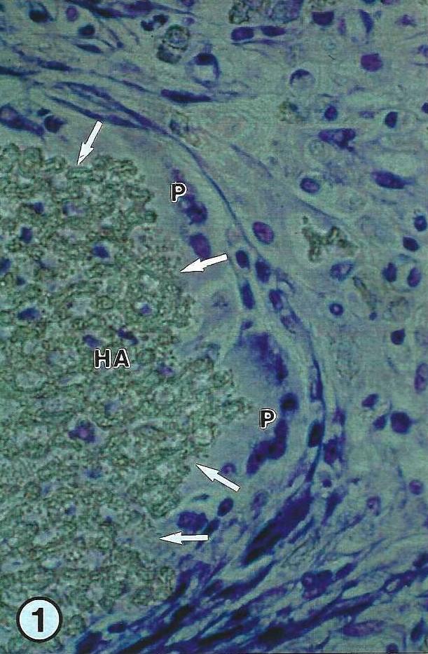
Using two types of HA (microporous and macroporous), the advantages of this method of histological and morphometrical examination will be demonstrated. The objective of the first study was to investigate, the suitability of a new porous HA (marine algae origin) to be used as an augmentation material for bone defects (Hotz et al., 1990). To serve as an augmentation material, the product should be absolutely resistant to resorption. Therefore, we studied the behavior of the biomaterial, after extraosseous implantation into paravertebral muscle pockets in rats. After one week, elliptical granules were enveloped by a young interparticulate soft tissue with a high vascularity. The biomaterial was progressively resorbed by mononuclear and polynuclear macrophages (Fig. 1). At higher magnification, polynuclear cells containing intact ceramic fragments (Fig. 2) were found.
The second portion investigated granular HA for its suitability as a carrier for bone morphogenetic protein (Urist et al., 1979). Allogeneic bone morphogenetic gelatin and macroporous HA were implanted into paravertebral muscle pockets in rats. After three weeks, remnants of the implanted bone gelatin were observed and they were bordered by both cartilage and by woven bone with active remodeling with osteoblasts and osteoclasts (Fig. 3). Appositional bone growth caused a direct physico-chemical binding with no soft tissue layer between the matrix induced heterotopic ossicles and the surrounding porous coralline HA (Fig. 4). Following labeling with fluorochromes (Rahn, 1976), the color bands confirmed appositional bone growth (Fig. 5).
|
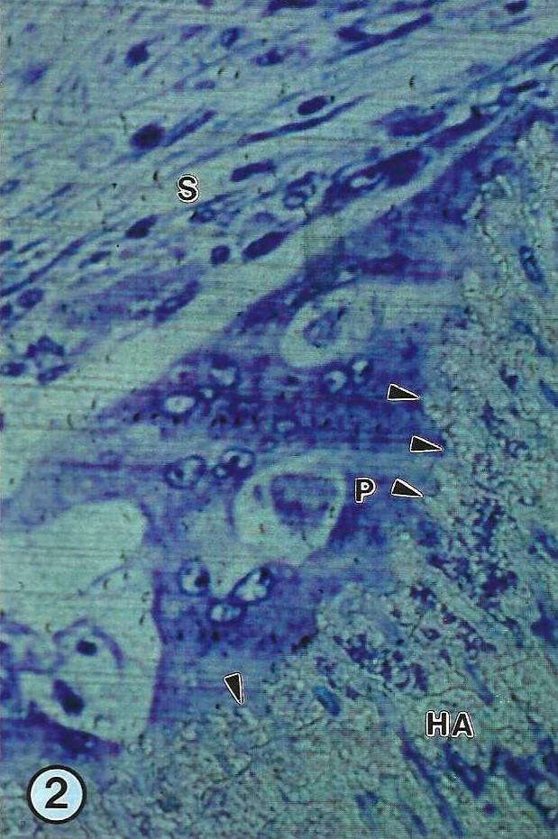 Figure 2. Higher magnification of a polynuclear cell (P) which lays with a "ruffled border like membrane system" on the surface (arrowheads) of the ceramic (HA). Soft tissue (S). X624. |
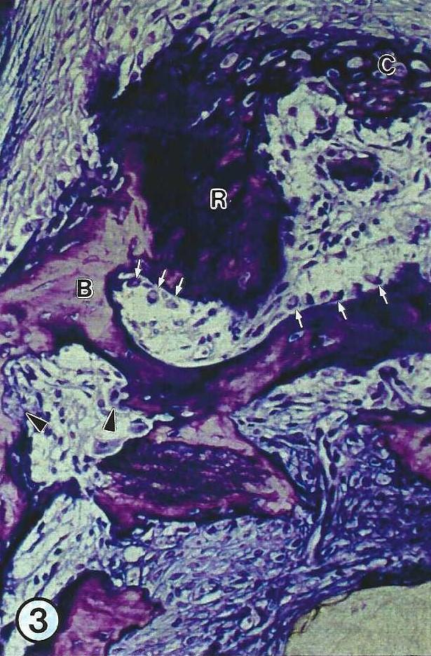 Figure 3. Remnants (R) of intramuscularly implanted bone morphogenetic gelatin (BMG) are bordered by cartilage (C) and by woven bone (B) with active remodeling with osteoblasts (arrows) and osteoclasts (arrowheads). Toluidine blue stain,. X64. |
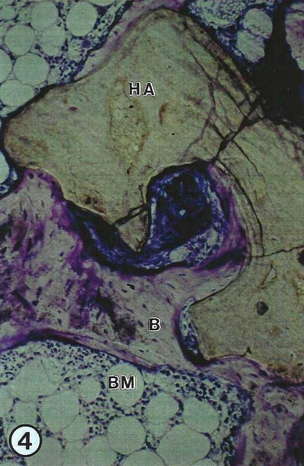 Figure 4. Appositional new bone formation around the ceramic with direct ceramo- osseous binding. Porous coralline hydroxylapatite (HA) (Interpore 200), Woven bone (B), Bone marrow (BM); Toluidine blue stain, X64. |
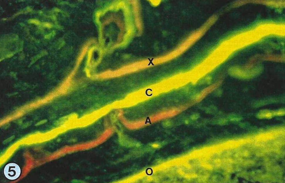 Figure 5. The color bands, following labeling with fluorochromes, indicate appositional bone growth [6 weeks: Xylenolorange (X); 7 weeks; Calcein ( C); 8 weeks; Alizarinkomplexon (A); 11 weeks: Oxytetracycline (0)]. X250. |
|
Donath K, G. Breuner: A method for the study of undecalcified bones and teeth with attached-tissues - the Sage-Schliff- technique. J Oral Path 11:318,1982.
https://doi.org/10.1111/j.1600-0714.1982.tb00172.x
Gross UM, V Strunz: Surface staining of sawed sections of undecalcified bone containing alloplastic implants. Stain Technol52(4):217,1977.
https://doi.org/10.3109/10520297709116778
Holmes RE, HK Hagler: Porous hydroxylapatite as a bone graft substitute in cranial reconstruction: A histometric study. Plast Reconstr Surg 81(5):662,1988.
https://doi.org/10.1097/00006534-198805000-00003
Hotz G, B Krempien, G Mall: Degradation of microporous phycogene hydroxylapatite after extraosseous implantation. In: Bioceramics, Vol 2 (Proceedings of the 2nd International Symposium on Ceramics in Medicine, September 1989), G Heimke (ed), Heidelberg, Germany. German Ceramic Society, Cologne, p. 71, 1990. ISBN 3-925543-06-6.
Rahn BA:Die polychrome Fluoreszenzmarkierung des Knochenbaus. Zeiss Information 22:36,1976.
Schultz M, R Drommer: Moglichkeiten der Präparateherstellung aus dem Gesichtsschädelbereich fur die makroskopische und mikroskopische Untersuchung unter Verwendung neuer Kunststofftechniken. Fortschr Kiefer Gesichts Chir 28:95,1983.
Urist MR, AJ Mikulski, A Lietze: Solubilized and insolubilized bone morphogenetic protein. Proc Natl Acad Sci 76:1828,1979.
https://doi.org/10.1073/pnas.76.4.1828
von Hagens G: Emulsifying resins for plastination. Praparator 25:43-50,1979a.
von Hagens G: Impregnation of soft biological specimens with thermosetting resins and elastomers. Anat Rec 194:247- 256,1979b.
https://doi.org/10.1002/ar.1091940206
von Hagens G: Heidelberg Plastination Folder: Collection of all technical leaflets for plastination. Anatomisches Institut 1, Universitat Heidelberg, 1985.