1 Department of Anatomy, Histology and Embryology, Semmelweis University, Faculty of Medicine. Tiizolt6 u.58., Budapest, Hungary, H- 1094.
2 Department of Pathology, Semmelweis University of Medicine, 01/oi u.93., Budapest, Hungary, H- 1094.
Numerous recent studies have acknowledged the merits of plastination in the anatomical sciences. Specimens conserved in this way can be handled easily and hygienically which broadens the possibilities in the presentation of anatomical preparations. The present study was aimed to introduce plastination in another morphological discipline, pathology. Conserving pathological organs or tissues with polymer impregnation and curing offer completely different challenges than when plastinating healthy tissues. Diseased tissues often have color and/or consistency changes which may be washed out or altered during preservation. However, preservation of these tissue changes in disease is of primary diagnostic importance. It can be demonstrated that color differences can be preserved in many cases as well as fixation of loosely anchored tissues without alteration of consistency is possible. Subtle, but typical alterations on the surface of diseased organs are demonstrable as well. At the same time, differences between neighboring healthy and pathological tissues can completely disappear which means a loss of important diagnostic cues. Regarding the large number and diverse kinds of plastinated specimens that have been processed, plastination serves as a useful tool in preserving pathological tissues.
plastination; pathology; tissue
Telephone: 36 1 21 56 920 /3600; Fax: 36 1 215 5158; E-mail: alpar@ana.sote.hu

![]()


Plastinated specimens were introduced to morphological disciplines more than twenty-five years ago and have proven to be of great use (von Hagens et al., 1 987). They are dry and hygienic which enables them to be used in an everyday environment (von Hagens et al., 1985). Our previous study along with numerous others reported that both instructors and students have found plastinated specimens as useful supplements in the anatomy curriculum (Mansor, 1996; Alpar et al., 2001; Latorre et al., 2004; Lozanoff, 2004; Riederer et al., 2004 ; Serodio et al., 2004 ; Latorre et al., 2006).
Recently, we have broadened our activity in plastination to include pathological specimens as have many institutions (Kularbkaew and Cook, 1996; Martin Alguacil and Martin-Orti, 2004). More than one hundred specimens have been plastinated in the Department of Anatomy, Histology and Embryology for the Department of Pathology, Semmelweis University of Medicine. Preservation of pathologic specimens has produced new challenges. Instead of the fixation of healthy organs, pathologically transformed tissues have to be preserved showing the characteristics which differ from those of normal tissues. Tumorous, inflamed, or degenerated tissues need to be plastinated with their typical surroundings preserved as well. In other cases, subtle color differences which were of primary importance in pathological diagnoses need to be retained.
Although plastinated specimens cannot replace the experience of dissection and the survey of fresh, unfixed organs, they can be used outside the dissection room. For this reason, they are useful supplements to student studies and in the preservation of precious rarities.
In order to investigate the conservation of different types of diseased tissues, specimens destined for plastination were selected from a broad range of diseases. Specimens were fixed in 4% formalin for a minimum of three weeks. Subsquently, they were dehydrated by freeze substitution with -25°C acetone. Four changes of acetone were carried out over an eight week period. After dehydration, specimens were impregnated in a reaction-mixture of SR10 polymer (Biodur™) with l% Catalyst SH03 (Biodur™) . Vacuum was adjusted to a slow rising of bubbles in the silicone. Bubbles lasted four weeks. The specimens were returned to atmospheric pressure, removed from the silicone bath, warmed to room temperature and prepared for curing with Gas Cure SH06 (Biodur™). In order to preserve the often rather vulnerable mutations, specimens were handled with care. Specimens were placed in a closed atmosphere, which was saturated by gas cure using an aquarium air pump working continuously for seven hours per day the first two days. Calcium chloride was used to control moisture in the curing chamber. Specimens were wiped every two hours the first day. Thereafter they were kept in the container for seven days and wiped as needed .
 Figure 1. Coronal section of human brain with blood filled lateral ventricles (A), longitudinal section through human heart and pericarial sac demonstrating cardiac tamponade with the causative lesion at arrow (B), longitudinal section of human descending aorta with ulcerated, hemorrhagic atherosclcrotic plaques at arrows (C), transverse section through human brain with intraparenchymal hemorrhage at arrows (D). |
Although significantly different in consistency, extensive brain hemorrhage which fully invaded the ventricles was plastinated preserving the typical location of the hemorrhage (Fig. l a). Similarly, pericardiac tamponade was also demonstrable along with the exact locus of the ventricular wall rupture (Fig. l b). The striking color of ulcerated, hemorrhagic atherosclerotic plaques was preserved after plastination in the wall of the descending aorta (Fig. l e). Preservation of color differences aided identifcation of intraparenchymal bleedings in the brain caused by multiform glioblastoma (Fig. I d).
The typical fine structure of the tumor was recognized on most specimens. The vortical, fibrous construction of an ovarian fibroma was visible (Fig. 2a) as was the spreading and osteolytic character of a bone metastasis (Fig. 2b).
Characteristic features, i .e. color or surface differences of inflamed tissues were preserved. The typical multiple pin shaped and sized microabscesses of purulent pyelonephritis were recognizable on the surface of the kidney (Fig. 3a). The yellow-green color of a purulent meningitis could also be demonstrated after plastination (Fig. 3b).
After plastination the characteristic fibrous, uneven surface of fibrinous pericarditis remained demonstrable (Fig. 4a). As well, erosions could also be observed on the mucosa of a metastatic infiltrated stomach (Fig. 4b).
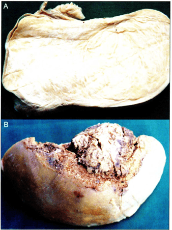 Figure 2. Longitudinally sectioned human ovary with ovarian fibroma (A ), osteolytic metastasis in the epiphyseal region of a bone (B). |
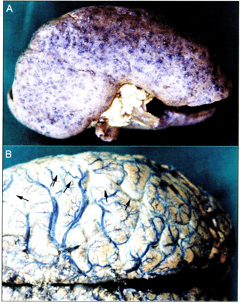 Figure 3. Plastinated kidney demonstrating purulent pyelonephritis (A). Plastinated brain (B) demonstrating purulent menengitis at arrows. |
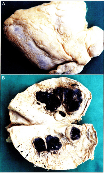 Figure 4. Subsinousal surface of plastinated human heart demonstrating fibrinous pericarditis (A), mural erosions (at arrows) in a metastatic infiltrated human heart ( B). |
During the last decade, the Department of Pathology at Semmel weis University of Medicine has collected a hundred specimens which represent outstanding examples of different pathological diseases or rarities in medicine. Tumors and other pathologies are fixed to healthy tissues which presents a challenge in collecting, as well as, preparing tissue for plastintion. In spite of these problems many pathological tissues have now been plastinated.
Numerous preparations of various diseases and organs were successfully plastinated which suggests that diverse types of pathological tissues can be properly conserved by this method. The present study offers insight into the possibilities in the plastination of pathological specimens. A great challange in preserving these specimens by plastination is the preservation of small, but diagnostically important colors and textures of the tissues.
Subtle changes on the surface of organs remained demonstrable irrespective of the kind of the mutation; degenerative (Fig. le), tumorous (Fig. 2b) and inflammatory (Fig. 3a) changes were equally recognizable. The inner structure of various diseased tissues could also be studied on the cut surface of the organ, typically in tumors which enabled the observer to identify both the type and the extension or demarcation.
Color differences are of primary importance in pathological diagnostics which can be properly studied only on fresh and unfixed specimens. On most plastinated preparates, the true colors were faded. This was due to formalin fixation and dehydraton but not to the silicone plastination process. Nevetherless, typical color differences could be properly demonstrated on numerous specimens (Figs. I a, I c, 3b).
Diagnostics also depend upon recognition of alterations in the consistency of the diseased tissue. This helpful feature is completely lost when the organ is plastinated which much be acknowledged as an inevitable shortcoming of the method. It has to be mentioned, however, that the consistency of both health and tumorous or degenerative tissues is changed during formalin fixation as well.
Fixation of non-stable tissues, e.g. bleeding (or hemhorrage) is another problem to be solved. In addition to the risk that the hemorrhage could be easily washed out, its shrinkage is different from that of the surrounding tissues. Still, hemorrhages could be plastinated in loco on severa l specimens (Fig. 1 A-C).
Considering the large number and variety of plastinated specimens, we suggest that plastination can be usefully applied in the conservation of pathological tissues.
Acknow ledgements
We would like to thank M. Toth and L. Patonay for the quality photos.
Alpar A, Ga l A, Patonay L, Kalman M . 2001 : Local flaps for fingertip injuries: a plastinated model. J Int Soc Plastination 16:42-45.
https://doi.org/10.56507/XOKS5488
Kularbkaew C, Cook P. 1996: Plastinated pathology specimens at room temperature in Thailand. J Int Soc Plastination 11 (1):17-19.
https://doi.org/10.56507/HNQI6175
Latorre R , Garcia-Sanz M P, Gil F, Moreno M , Agut A, Quinonero JM, Lozano E, Herrero J , Hernandez- Pina F, Fenandes-Serodio H , Henry R. 2004: Evaluation of plastinated organs as a resource for improvement of the teaching-learning processes. Abstract presented at The 12th International Conference on Plastination, Murcia, Spain July 11-16, 2004 . J Int Soc Plastination 19:48.
Lozanoff S. 2004 : Plastination : A tool for education. Abstract presented at The 12th International Conference on Plastination, Murcia, Spain July 11-16, 2004. J Int Soc Plastination 19 (1): 11.
Mansor 0. 1996: Use of plastinated specimens in a medical school with a fully integrated curriculum . J Int Soc Plastination 11(1): 16-17
https://doi.org/10.56507/MMLW7935
Martin-Alguacil N , Martin-Orti R. 2004: Plastination of a canine encephalon with hydrocephalus. Abstract presented at The 12th International Conference on Plastination, Murcia, Spain July 11-16, 2004. J Int Soc Plastination 19:59.
Riederer BM , Musumeci E, Duvoisin B, Lang FJ W . 2004: Plastination , a useful tool in teaching clinical anatomy. Abstract presented at The 12th International Conference on Plastination, Murcia, Spain July 11-16, 2004. J Int Soc Plastination 19:43.
Serodio HC, Gouvviea J, Lameiras JM, Gil F, Rameriz G, Latorre R. 2004: Plastination and animation: Pedagogic-didactic proposal for study of fetal circulation in ruminant fetuses, as a paradigm for the study in other mammals . Abstract presented at The 12th International Conference on Plastination, Murcia, Spain July 11-16, 2004. J lnt Soc Plastination 19:52 .
von Hagens G. 1985: Heidelberg Plastination Folder : Collection of all technical leaflets for plastination. Heidelberg , Germany: Anatomisches Institut I, Universitiit Heidelberg.
von Hagens G, Tiedman K, Kriz W. 1987 : The current potential of plastination . Anat Embryol 175(4):41 1 - 421.
https://doi.org/10.1007/BF00309677