Royal Veterinary College, London, United Kingdom, NW1 0TU
Embalming and plastination are both processes which tend to reduce the amount of natural color within a specimen. This can result in a lack of definition between tissue types making them less effective as teaching resources. The Mazwell Group© manufactures and distributes Dodge© embalming chemicals for the funeral industry which are intended to give a life-like appearance. The Royal Veterinary College (RVC) has begun to use these chemicals as standard for their anatomical embalming. However, there is conflicting literature on whether these types of chemicals are suitable precursors to plastination, a process which the RVC also carries out. Bovine, canine, and equine hearts were plastinated following removal from cadavers embalmed using Dodge© embalming chemicals. The final specimens were compared both visually and with an RGB color sampler, with those plastinated following embalming with a standard formalin/water/glycerol solution. Comparisons of the heart specimens and others showed an increase in color retention and tissue differentiation. The use of Dodge© embalming chemicals has been shown not to be detrimental to the plastination process and results in specimens suitable for anatomical teaching.
anatomy; embalming; hearts; plastination; veterinary
Sarah Nicoll, Royal Veterinary College, Department of Comparative Biomedical Sciences, 4 Royal College Street, London, NW1 0TU, UK. Tel: 02039054636 Email: snicoll@rvc.ac.uk

![]()


Embalming is an ancient process that has evolved considerably through time. Today, it is not only valuable as a part of the funeral industry, but also as a part of medical and veterinary anatomical education. Plastination, by contrast, is a relatively modern process, having been developed in the late 20th century (Henry et al., 2019), and is still evolving. Embalming and plastination are both methods of preservation, vastly different in their processes, but one is often necessary for the other to take place. The Royal Veterinary College (RVC) has a plastination laboratory which produces a wide range of specimens for teaching and widening participation activities. The system in use at the RVC is the cold-temperature silicone standard S10 technique, and requires the specimens to be embalmed in the first instance (Henry et al., 2019).
In the last few years, the RVC has transitioned from using a simple formalin/water/glycerol (FWG) solution to using embalming chemicals originally intended for the funeral industry, the reason being that they improved upon the primary criteria of longevity, color retention and ease of specimen storage. The Mazwell Group©, which produces the Dodge© embalming chemicals (The Mazwell Group 2020a) now used at the RVC, has a wide range which can be mixed to achieve the desired results. One of the chemicals selected from the range, Introfiant (The Mazwell Group 2020b), contains a dark reddish/pink stain called Dynachrome which gives the cadavers a natural appearance in terms of color. When the RVC transitioned to Dodge© embalming chemicals the outcome of how they would perform in conjunction with the plastination process was unknown. This technical report will describe: the chemicals and the process used to embalm canine, equine, and bovine cadavers; the process used to plastinate the isolated hearts taken from the embalmed canine, equine and bovine cadavers; and the results following plastination.
Embalming
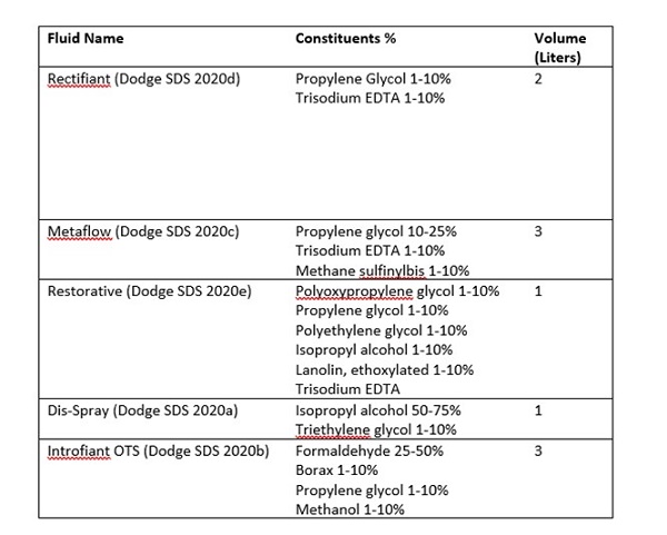 Table 1: The Dodge© embalming chemicals making up the Royal Veterinary College’s 10 Liter solution |
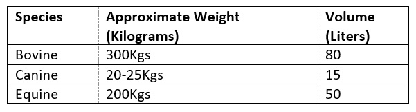 Table 2: The volume of embalming solution used in each species |
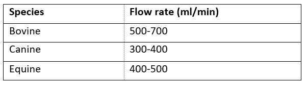 Table 3: Flow rate of embalming solution used for each species during embalming |
Three cadavers were used in this study: bovine, canine, and equine. All three were sourced in accordance with the RVC’s ethical guidelines. The chemicals used to embalm these cadavers are described in Table 1. The embalming solution is made up in batches of 10 liters.
Each cadaver used a different volume of the solution, depending on the species and size (Table 2). The Dodge© embalming machine (The Mazwell Group 2020c) has the ability to vary pressure and flow rate, and select between pulse and continuous flow, all of which are important when embalming cadavers of varying sizes.
The cadavers were cannulated at the carotid artery, and the embalming solution was introduced at a pressure of approximately 965 kPa (140 psi). The flow rate chosen depended on the size of the cadaver (Table 3). A drain was placed in the jugular vein allowing the flushing out of blood. The drain was closed when the majority of the fluid passing through the drain was embalming solution.
The canine cadaver was embalmed over approximately 3 hours. The equine and bovine cadavers took 6-8 hours.
Embalming was considered to be complete when tissues felt fuller to the touch. There was some firming of the tissues, but this effect mostly appeared over the hours/days following embalming. Superficial veins were distended, and there was visible staining of the mucous membranes. The skin was also stained where this was visible, depending on thickness, original color, and hair coverage. Following embalming, the drains and cannulas were removed. The cadavers were washed, wrapped/bagged, and stored in a cold room maintained at approximately 2-5° C for a minimum of 3 weeks before they were used for dissection/prosection.
Following the completion of teaching with the embalmed cadavers, the hearts were all selected as being good enough to retain for plastination. All unwanted tissue was removed from the specimens. They were then sectioned and returned to temporary storage prior to plastination.
Plastination
The cold-temperature silicone standard (S10) technique is the method employed by the RVC. Prior to plastination, the specimens were washed in room-temperature running water for a minimum of 24 hours to remove surface embalming solution and loose tissue or debris. The stages of plastination used for these heart specimens were as follows:
Dehydration
The specimens were immersed in acetone within stainless steel containers inside a freezer at a temperature of approximately -20°C. The acetone purity was regularly measured at 20°C using an acetonometer. The acetone was changed to increase the concentration surrounding and within the specimens. Acetone measurements and changes took place on average once a week. Dehydration was considered to be complete when readings were maintained in excess of 98.5% acetone.
Silicone Impregnation
The specimens were transferred to a vacuum chamber and submerged in the liquid silicone (S10+S3) solution. This vacuum chamber resides within a freezer maintained at -20°C, and is connected to a pump. A vacuum was slowly applied to the vacuum chamber with the silicone solution monitored for acetone bubbles. The system at the RVC does not use a manometer. Whenever acetone bubbles subsided, the vacuum was adjusted very slowly using the taps, until bubbles resumed. This was repeated until all taps were closed and all acetone bubbles had ceased rising from the silicone solution. At this point silicone impregnation was considered complete. Impregnation of these specimens took 4-5 months. This is longer than it might have been, however, these specimens were not processed in isolation, being mixed with specimens of different sizes and tissue types. Once impregnation was completed, the vacuum pump was turned off, and the taps opened allowing air to re-enter the vacuum chamber. The specimens remained in the vacuum chamber for a minimum of 24 hours before removing onto drip trays.
The specimens were allowed to rest on drip trays for several days. They were frequently rotated to allow excess silicone to drain from them and were also regularly wiped. When the amount of silicone dripping or exuding from the specimens had diminished considerably, the specimens were ready for curing.
Curing
The specimens were transferred to a curing chamber, where they were exposed to a vaporized curing solution (S6). They were rotated and wiped on a daily basis, to prevent the curing of any remaining pools or drips of silicone on the surface of the specimens. The specimens remained in the curing chamber for several days. Once they were no longer obviously tacky to the touch, the specimens were transferred on to paper towels for several days to check that they did not return to being tacky. If at any point they resumed being tacky, they were returned to the curing chamber for a few more days. Once the specimens had remained dry for several consecutive days, curing was considered complete. Total curing time varied between specimens, with the dog heart curing in approximately 1 week, and the equine and bovine hearts approximately 3 weeks.
Imaging
All photographs of specimens taken for this study were carried out on the same day with the same lighting, background, and camera.
Color Analysis
The colors of the plastinated Dodge© embalmed specimens were compared with those of previous plastinated specimens that had been embalmed with an FWG solution. The colors were sampled using an online RGB color tester (Rapid Tables 2021) which provided the RGB and #HEX values of each color, as well as a basic color name. Photographs of two specimens for comparison were pasted into the color picker. Several regions and tissue types were sampled, with three readings taken in each area to ensure variations in the colors across tissues were accounted for. Wherever possible, areas sampled were those in direct light rather than those in shadow.
The final plastinated heart specimens can be seen in Figures 1-6.
 Figure 1. Plastinated bovine heart, sectioned surfaces. Embalmed with Dodge© chemicals |
 Figure 2. Plastinated bovine heart, outer surface. Embalmed with Dodge© chemicals |
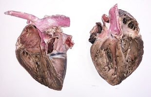 Figure 3. Plastinated canine heart, sectioned surfaces. Embalmed with Dodge© chemicals |
 Figure 4. Plastinated canine heart, outer surfaces. Embalmed with Dodge© chemicals |
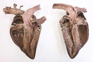 Figure 5. Plastinated equine heart, sectioned surface. Embalmed with Dodge© chemicals |
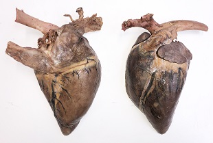 Figure 6. Plastinated equine heart, outer surface. Embalmed with Dodge© chemicals |
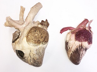 Figure 7. Plastinated bovine hearts, outer surfaces. The specimen on the left was embalmed with a traditional formalin/glycerol/water solution. The specimen on the right was embalmed with Dodge© chemicals |
 Figure 8. Plastinated canine hearts, outer surfaces. The specimen on the left was embalmed with a traditional formalin/glycerol/water solution. The specimen on the right was embalmed with Dodge© chemicals |
Examples of hearts plastinated following the RVC’s previous FWG embalming solution can be seen alongside those embalmed with the Dodge© chemicals in Figures 7 and 8.
The colors of the bovine and canine specimens shown in Figures 7 and 8 were sampled using the Image Color Picker (Rapid Table 2021). There were no plastinated FWG embalmed equine specimens available for comparison.
Table 4 shows the color sampling of four tissue types across both plastinated bovine hearts (Figure 7). Three samples were taken in each area, and the median RGB and color name for each region was calculated. The FWG embalmed bovine heart had colors in the grey and silver categories, whereas the Dodge© embalmed bovine heart had colors in the maroon, purple, grey, and white categories. Table 5 shows the color sampling of three tissue types across both canine plastinated hearts (Fig. 8). Again, three samples were taken in each area, and the median RGB and color name for each was calculated. The FWG embalmed canine heart was found to have colors which were categorized as variations of grey across all tissue types. The Dodge© embalmed canine heart was found to have a combination of grey and purple colors across the three tissue types sampled.
 Table 4: RGB Analysis comparing of the Dodge© embalmed plastinated bovine heart with a formalin/water/glycerol embalmed plastinated bovine heart |
 Table 5: RGB Analysis comparing of the Dodge© embalmed plastinated canine heart with a formalin/water/glycerol embalmed plastinated canine heart |
Analysis of the colors of these specimens showed that the FWG embalmed specimens have colors within the grey and silver spectrum, whilst the Dodge© embalmed specimens seem to have a wider range of colors across tissue types.
Even without the RGB color analysis it can be easily seen in Figures 1-8 that the heart specimens embalmed with the Dodge© chemicals have, to varying degrees, retained the artificial color introduced at the embalming stage. The bovine heart appears to have retained the most color, and the equine heart the least. The reasons for this variation in color retention across the three examples is unknown. Visual comparisons were also made with plastinated specimens from different body regions and tissue types (Figs. 9-11), embalmed with the Dodge© and FWG solutions. These also demonstrated a similar trend, with the Dodge© embalmed specimens exhibiting a wider range of colors.
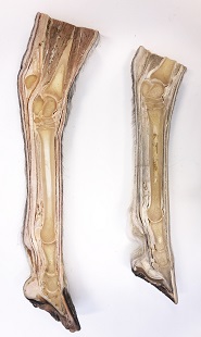 Figure 9. Plastinated equine distal hindlimb. The specimen on the left was embalmed with Dodge© chemicals. The specimen on the right was embalmed with a traditional formalin/glycerol/water solution |
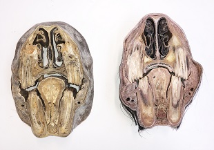 Figure 10. Plastinated transverse section of the equine head at the level of the nasal cavity. The specimen on the left was embalmed with a traditional formalin/glycerol/water solution. The specimen on the right was embalmed with Dodge© chemicals |
 Figure 10. Plastinated transverse section of the equine head at the level of the nasal cavity. The specimen on the left was embalmed with a traditional formalin/glycerol/water solution. The specimen on the right was embalmed with Dodge© chemicals |
When all these specimens were photographed, they were at least 18 months post-curing, some considerably more so. There appeared to be no deterioration in the Dodge© specimens, including any obvious changes in color or shape. There was a small amount of white dust accumulated on the surface of two of the specimens, visible in Figures 1 and 6, but this was easily wiped away with a dry piece of tissue.
An unexpected result of the process described was that, during dehydration of the Dodge© embalmed specimens, it was observed that the acetone leached some of the red stain from the specimens. This did not appear to have any detrimental effect on the process itself, or the final result. It was, however, also observed that other specimens embalmed with just formalin/water/glycerol that went through the dehydration process at the same time, seemed to take on a little of the red stain. Again, this did not appear detrimental to these specimens and may have even improved upon their appearance.
Traditional embalming solutions used for specimens intended for plastination usually include formalin at 5-15% (Henry et al., 2019), however, higher concentrations of 10-20% should be used for brains (Henry et al., 1997). Based on the constituent concentrations shown in Table 1, the RVC’s embalming solution formalin concentration ranges between 7.5% - 15%. Whichever concentration of formalin chosen, there are differing opinions on whether the addition of further chemicals to the embalming solution is beneficial or disruptive to the plastination process. Some believe embalming solutions containing alcohols, glycerin, glycols and/or phenol should not be used on specimens destined for plastination (DeJong and Henry, 2007). Others, however, have used solutions containing one or more of these chemicals and are satisfied with the results (Cook and Dawson, 1996; Pretorius, 1996; Norman and Nicoll, 2017).
Some authors have gone down the route of having two separate embalming solutions, one for dissection specimens, and one specifically for specimens intended for plastination (Cook and Dawson, 1996). This would not be a suitable option for the RVC, as the majority of the specimens plastinated are those which are initially either used as student dissection specimens during undergraduate teaching, or as prosections produced by staff for demonstration. Therefore, the embalming chemicals selected by the RVC are primarily chosen to provide teaching specimens which are not only long lasting and easy to store, but also provide good color and tissue differentiation during dissection. Good quality student dissection or prosection specimens may then be retained once finished with, for plastination. Only a very small number of RVC specimens are ever embalmed with plastination being the primary goal.
Most embalming solutions are traditionally transparent, which, when used in conjunction with the standard embalming process of draining blood from the cadaver, can produce a very pale grey/beige-looking specimen. This is not ideal, as good tissue differentiation is key in aiding student identification and understanding of anatomical structures. In the past, various embalming solutions were developed to aid the preservation of color e.g., Kaiserling’s, Klotz, Jores’, and McCormick’s solutions, and other coloring chemicals, such as eosin and merthiolate (Iliff et al., 2019). However, these seemed to yield mixed results. Plastination can further bleach the color from specimens during the dehydration stage (McCreary et al., 2013) leaving specimens pale, with features harder to distinguish. Today, in order to achieve a more natural and useful appearance, many plastinated specimens are painted or stained (Raoof et al., 2013; Mccreary et al., 2013; Yu et al., 2014; Kang et al., 2015). A stain could be added to the acetone bath, or a pigmented silicone could be used during the impregnation stage of plastination. These options are unfortunately not selective, so all the tissues will be the same color (Iliff et al., 2019), which would not be an improvement. The ideal would be to have an embalming solution which produces good tissue color differentiation upon dissection, which is also suitable for plastination.
This study has looked at the production of plastinated specimens using a colored embalming solution, however the number of specimens used was limited and further investigations should be made into its wider suitability.
Cook P, Dawson B. 1996: An improved method of embalming suited to subsequent plastination requirements. J Int Soc Plastination 10:34
https://doi.org/10.56507/AHUU1684
DeJong K, Henry RW. 2007: Silicon plastination of biological tissue: cold-temperature technique biodur technique and products. J Int Soc Plastination 22:2-14.
https://doi.org/10.56507/ZLMJ7068
Dodge SDS. 2020a: Dis-Spray Safety - Data Sheet. URL: https://shop.dodgeco.com/content/files/SDS/DisSpray.pdf [Accessed April 2020]
Dodge SDS. 2020b: Introfiant OTC - Safety Data Sheet. URL: https://shop.dodgeco.com/content/files/SDS/IntrofiantOTC.pdf [Accessed April 2020]
Dodge SDS. 2020c: Metaflow - Safety Data Sheet. URL: https://shop.dodgeco.com/content/files/SDS/Metaflow.pdf [Accessed April 2020]
Dodge SDS. 2020d: Rectifiant - Safety Data Sheet. URL: https://shop.dodgeco.com/content/files/SDS/Rectifiant.pdf [Accessed April 2020]
Dodge SDS. 2020e: Restorative - Safety Data Sheet. URL: https://shop.dodgeco.com/content/files/SDS/Restorative.pdf [Accessed April 2020]
Henry RW, von Hagens G, Seamans G. 2019: Cold temperature/Biodur®/S10/von Hagens'-Silicone plastination technique. Anat Histol Embryol 48(6):532-538.
https://doi.org/10.1111/ahe.12472
Henry RW, Janick L, Henry C. 1997: Specimen preparation for silicone plastination. J Int Soc Plastination 12(1):13-17.
https://doi.org/10.56507/HVSK9838
Iliff S, Concha I, Chereminskiy V, Henry RW. 2019: Coloring plastinated specimens, Anat Histol Embryol 48(6):552-556.
https://doi.org/10.1111/ahe.12506
Kang J, Lliff S, Henry RW, Hermey D. 2015: Coloring muscles and vessels of plastinated limbs with colored silicone to supplement teaching. J Plastination 27(2):9-12
https://doi.org/10.56507/LTNC5138
McCreary J, Iliff S, Herney D, McCreary K, Henry RW. 2013: Silicone-based coloration technique developed to highlight plastinated specimens, J Plastination 25(2):13-20.
https://doi.org/10.56507/XLBR3803
Norman RT, Nicoll S. 2017: Creating a teaching aid for ferret reproductive and adrenal anatomy. J Inst Anat Sci 18:43-47.
Pretorius WF. 1996: Formula for embalming of cadvers for student dissection and the modification thereof for plastination. J Int Soc Plastination 10:35-36.
https://doi.org/10.56507/ABGU2279
Raoof A, Marchese C, Marchese LA, Falk KC, Mirafzali N. 2013: Painting plastinated neurovascular pathways : evaluation of coloring techniques. J Plastination 25(2):21-26.
https://doi.org/10.56507/LJZQ6496
Rapid Tables. 2021: Image Color Picker. URL: https://www.rapidtables.com/web/color/color-tester.html [Accessed April 2021]
The Mazwell Group. 2020a: Our Journay. URL: https://www.themazwellgroup.com/about-us [Accessed April 2021]
The Mazwell Group. 2020b: Introfiant. URL: https://www.themazwellgroup.com/product-page/introfiant [Accessed April 2021]
The Mazwell Group. 2020c: The Original Dodge Embalming Machine. URL: https://www.themazwellgroup.com/product-page/the-original-dodge-embalming-machine [Accessed April 2021]
Yu S-B, Zhang J-F, Chi Y-Y, Gao H-B, Liu J, Sui H-J. 2014: Plastination of a whole horse for veterinary education. J Plastination 26(1):29-32.
https://doi.org/10.56507/UKKJ2013