'School of Physiotherapy, Department of Orthopedic Surgery and Physiotherapy, Faculty of Medicine Siriraj Hospital, Thailand.
²Department of Anatomy, Faculty of Science, Mahidol University, Bangkok, Thailand.
Human brain slices, 4-6 mm thick, were stained and plastinated. Five methods of staining were compared: Mulligan's, Le Masurier's, Roberta's, Braak's and Alston's methods. The gray matter stained grayish black, brilliant blue, reddish brown, bluish green and brickred, respectively. Shrinkage after staining was 2% in Braak's method and less than 1 % in the other methods. After plastination, the shrinkage was approximately 10% in all methods. Judging from staining cost, time spent and contrast between the gray and white matter, Alston's method was the best among the five studied. All stained, plastinated brain slices displayed anatomical details suitable for neuroanatomy study.
Brain slices, Neuroanatomy Teaching
Boonsirm Withyachumnarnkul, M. D. Ph. D., Associate Professor, Department of Anatomy, Faculty of Science, Mahidol Uni- versity, Rama 6 Rd., Bangkok 10400, Thailand. Telephone: 246 1360 78 Ext. 7777 / Fax: 246 7056. Email: scbwt@mahidol.ac.th

![]()


Brain slices can be plastinated by both conventional P40 and S10 techniques (von Hagens, 1979; von Hagens, 1985/86; Ulfig and Wuttke, 1990). The disadvantage of the P40 technique, from our personal observation, is that the resultant semi-transparent plastinated brain slices require a light box to optimally visualize anatomical details. Brain slices plastinated by the S10 technique, however, require macroscopic staining to differentiate between the fiber tracts and neuronal components. When Ulfig and Wuttke (1990) stained brain slices with Astra blue followed by plastination, the finished product displayed good contrast between the fiber tracts and neuronal components, without using the light box. The latter specimens were also durable and convenient to handle. In the present study, brain slices were stained with different methods and plastinated using the S10 tech- nique. The purpose was to find the most suitable method to prepare stained, plastinated brain slices for neuroanatomy instruction.
Brain Specimens and Sectioning
Human brains were obtained at post-mortem from autopsy rooms at Chiengmai Hospital, Ramathibodi Hospital and Siriraj Hospital; all are associated with medical schools in Thailand. The patients died from non-neurological causes. The brains were fixed in 4% formalin for six months. Brains from 60 year-old or older subjects stain poorly (Roberts and Hanaway, 1969) and thus were excluded from this study.
The fixed brains were washed in running tap water over- night before being serially sectioned with a meet slicer. Brains were sectioned in three planes: coronal (4 mm), horizontal (6 mm) and sagittal (6 mm). The sections were laid on wet filter paper and stacked individually on stainless steel mesh trays. The trays containing series of brain sections were secured and the whole assembly was placed in 4% formalin solution for an additional eight hours, after which brain slices were washed with running tap water overnight before staining.
Staining
Five methods of staining were carried out as follows:
Mulligan's Method (Gregg, 1975)
C . I min in 0.4% tannic acid (W/V in water) at room temperature
Le Masurier's Method or Prussian Blue Reaction's Method (Le Masurier, 1935)
Roberts's Method (Roberts and Hanaway, 1969)
Braak's Method or Astra Blue's Method (Braak, 1978)
Alston's Method (Alston. 1981)
*Polyclens is a proprietary paint remover and also a powerful lipid solvent. It was purchased from Polycell Holding Ltd, Welwyn Garden City, Herts, UK.
The staining procedures in all methods, especially the Braak's method, were carried out in a ventilation hood. For- mic acid used in the Braak's method produced offending fume that could be hazardous.
Plastination Procedure
Standard procedure for S10 plastination (von Hagens, 1985/86) was employed with slight modifications. The stained brain slices were dehydrated in 100% acetone at -25 °C for 2 days, with daily change of fresh acetone. The acetone: brain volume ratio was 10:1. The dehydrated specimens were immersed in S10/S3 (99:1 by volume) mixture in a plastination kettle for 2 days. Forced impregnation was started at the third day when the vacuum pump was turned on. Acetone bubbles were observed through the top glass- cover of the kettle and carefully adjusted; it took about two weeks to complete the impregnation step. Curing was accomplished by S6, with frequent wiping of the surface of the brain slices to prevent accumulated plastic on the sur- face.
Photographs were taken at each step after sectioning, after staining and after plastination, to calculate the degree of shrinkage following each step. A ruler was photographed with the brain slice at each step and two points of reference were selected from the photograph. A line was drawn be- tween these two points and the degree of shrinkage was calculated by comparing the length of the line in the non- stained, stained and plastinated brain slices.
The plastinated brain slices were framed with two clear, rectangular, plastic plates, for protection and convenient handling.
The finished brain slices are shown in figure 1.
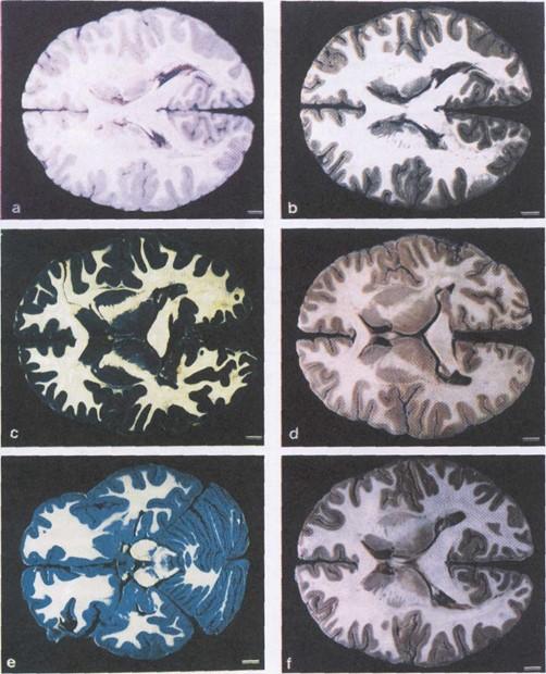 Figure 1. Comparison of plastinated brain slices, after staining with different methods, a - unstained brain section, b - Mulligan's method, c - Le Masurier's method, d - Roberts's method, e -Braak's method, f - Alston's method. Bar = 1 cm. |
Comparison between colors, percent shrinkage, time spent and staining cost is shown in table 1. All considered, the Alston's method was the best since it gave good contrast between the fiber and neuronal components, required minimum staining time and was economic. The staining step caused slight shrinkage of the specimens.
| Mulligan | Le Masurier | Roberts | Braak | Alston | |
| Color of Gray Matter | grayish black | brilliant blue | reddish brown | bluish green | brick-red |
| Color of White Matter | white | white | white | white | white |
| Shrinkage After Staining (%) | 0.6 | 0.7 | 0.6 | 2.0 | 0.4 |
| Shrinkage After Plastination (%) | 9.0 | 9.0 | 8.2 | 10.0 | 8.0 |
| Staining Time | 7 min | 13 min | 12 min | 2 days | 25 min |
| Staining Cost per Slice (US$) | 0.4 | 0.4 | 0.4 | 2.8 | 0.6 |
The Braak's method gave a slightly higher degree of shrinkage (2%) than the others (less than 1%). The shrinkage after plastination, approximately 10% in all methods, was more pronounced in the gray matter than in the white matter.
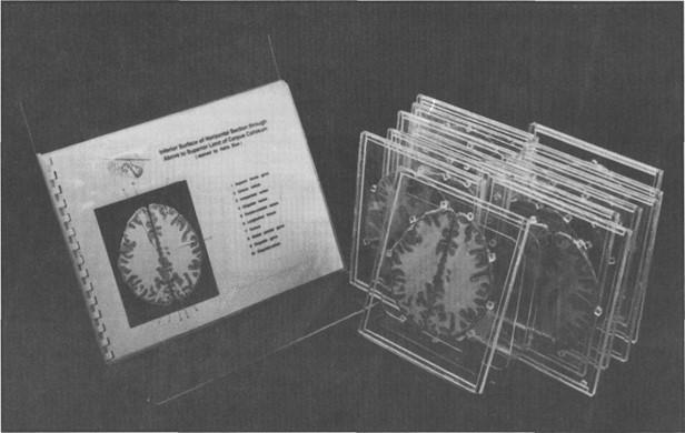 Figure 2. The plastinated brain slices after framing |
After plastination, the color was more intense and has remained stable for more than one year. After framing, the brain slices were convenient to handle and study (Figure 2). Their anatomical details including nerve tracts and nuclei were much clearer than fresh brain specimens. Total time for plastination, excluding the fixation period, was approximately one month (table 2)
| Fixation | > 6 months |
| Sectioning and Staining | 3 days |
| Dehydration | 5 days |
| Forced Impregnation | 2 weeks |
| Curing | 2 days |
| Framing | < l day |
| TOTAL | 7 months |
This study suggests that Alston's method for brain slice staining is the best among the five being tried. Although contrast between the fiber and neuronal components were best achieved in the Braak's and Alston's methods, the later, however, was superior to the former in several aspects.
Firstly, the cost of staining in Alston's method was about 4.5 x less than that of the Braak's method (table 1). Secondly, several brain slices could be stained at the same time in the Alston's method; while only a few slices at a time could be stained in the Braak's method, that also required constant shaking. Thirdly, the staining solution of the Alston's method could be re-used, while that of the Braak's method could be used only once. Fourthly, the degree of shrinkage after staining, and also after plastination, was lower in Alston's method (table 1). And, finally, toxic fume of formic acid used in the Braak's method was a potential hazard even being performed in the ventilation hood.
The mechanism underlying the development of colors by the staining procedure is still controversial. Mainland (1928) and Blair et al. (1932) suggested that the color development was due to the difference between the texture of the gray and white matters and different penetrability of the chemicals into the brain tissues. The latter might be due to the fact that the white matter contains more lipid (myelin sheath) than the gray matter. By applying Mulligan's solution, phenol dissolved lipid materials of the white matter to produce a jelly coat covering its surface. If the white matter was damaged, the differentiation between the gray and white matter (the damaged areas) would not be distinct (Main- land, 1928; Blair et al., 1932). Good preservation of the brain is thus very important in order to reveal the anatomical de- tails (Roberts and Hanaway, 1969). In Le Masurier's method, Sincke (1926) suggested that the color in Prussian blue re- action was developed from a chemical reaction between the staining agents and iron molecules present in the brain tis- sue. In Mulligan's method, a reaction of tannic acid with protein in the gray matter is proposed (Mulligan, 1931). Tannic acid might also react with polysaccharides which is rich in the brain tissue (Szabo and Roboz-Einstein, 1962; Sannes et al., 1978). Alston (1981) suggested that cupric sulfate in the Mulligan's solution probably interacted with the surface of the gray matter and potassium ferrocyanide then reacted with cupric sulfate to form cupric ferrocyanide.
The color developed in the Braak's method is most likely accomplished by a mechanism different than that of the Mulligan-related methods. The Astra blue, a mucopolysaccharide staining agent used in this method (Pioch, 1957; Fasske, 1957), probably stained mucopolysaccharide-rich area of the gray matter. Performic acid, also used in the method, reacts with cystine, tryptophan and methionine (Toennies et al., 1942). Cysteic acid was derived from oxidation of cystine by performic acid (Pearse, 1951; Lillie, 1952; Adams and Sloper, 1956). The concentration of cystine is increased in the gray matter during the later stage of myelination (Friede, 1966). The technique, however, took a relatively long period (2 days) and performic acid is toxic and difficult to handle.
The finding that the degree of shrinkage after plastination is more pronounced in the gray matter is probably due to the higher amount of water in the gray matter (82%) than in the white matter (72%) (Suzuki, 1981). The degree of shrinkage at approximately 10% is considered acceptable and did not cause any problems in learning neuroanatomy from these specimens.
Acknowledgment
The study was supported by the National Research Council, Thailand, 1993-94. The authors thank Professor Bhuket Wajanon and Associate Professor Sanjai Saengwichien for their constructive criticism and encouragement.
Adams CWM, Sloper JC: The hypothalamic elaboration of posterior pituitary principles in man, the rat and dog. Histochemical derived from a performic acid alcian blue reaction for cystine. J Endocrinol 13: 221-228, 1956.
https://doi.org/10.1677/joe.0.0130221
Alston RL: A batch staining method for brain slices allowing volume measurements of gray and white matter using an image analyzing computer (Quantimet 720). Stain Technol 56: 207-213, 1981.
https://doi.org/10.3109/10520298109067313
Blair DM, Davies F, McClelland EW: On the nature of certain macroscopic staining of the brain. JAnat 66: 478-485,1932.
Braak H: Simple and durable staining of thick sections of the human brain for macroscopic study. Stain Technol 53: 87-88,1978.
https://doi.org/10.3109/10520297809111447
Fasske E:1957. Zur Darstellung der sauren Mukopolysaccharide im Bereich der Zwischensubstanz mesenchymaler Gewebe. Zbl Allg Path Anat 97: 174- 179,1957.
Friede RL: Proteins and Amino Acids. In: Topographic Brain Chemistry. Edited by Friede RL, New York: Academic Press, pp 357-387,1966.
Gregg RV: Tannic acid-iron alum reaction: Stain of choice for macroscopic sections of brain to be embedded in plastic. Stain Technol 50: 87-92,1975.
https://doi.org/10.3109/10520297509117040
Le Masurier HE: Simple method of staining macroscopic brain sections. Arch Neurol Psychiat 34: 1065-1067, 1935.
https://doi.org/10.1001/archneurpsyc.1935.02250230137011
Lillie RD: Ethlenic reaction of ceroid with performic acid and Schiff reagent. Stain Technol 27: 37-45,1952.
https://doi.org/10.3109/10520295209105058
Mainland D: Uber makroskopische Farbung von Gehirnpraparaten mit Berlinerblau. Anat Anz 65: 84- 88,1928.
Mulligan JH: Amethod of staining the brain for macroscopic study. JAnat 65: 468-472,1931.
Pearse AGE: The histochemical demonstration of keratin by methods involving selective oxidation. Q J Micr Sci 92: 393-402,1951.
https://doi.org/10.1242/jcs.s3-92.20.393
Pioch W: Uber die Darstellung saurer Mucopolysaccharide mit dem Kupferphthalocyaninfarbstoff Astrablau. Vir- chow's Arch Path Anat 330: 337-346,1957.
https://doi.org/10.1007/BF00954964
Roberts M, Hanaway J: Preparation of brain slices for macroscopic study by the copper sulfate-phenol- ferrocyanide technique. Stain Technol 44: 143-146, 1969.
https://doi.org/10.3109/10520296909063340
Sannes PL, KatsugamaT, Spicer SS: Tannic acid-metal salt sequences for light and electron microscopic localiza- tion of complex carbohydrates. J Histochem Cytochem 26: 55-61,1978.
https://doi.org/10.1177/26.1.74385
Sincke G: Eine neue Methode zur Farbung von makroskopischen Gehirn schnitten. Anat Anz 61:311- 313,1926.
Suzuki K: Chemistry and Metabolism of Brain Lipids. In: Basic Neurochemistry. Edited by Siegel GJ, Alters RW, Agranoff BW and Katzman R. 3rd., Boston: Ed. Little Brown and Company, pp 355-370,1981. .
Szabo MM, Roboz-Einstein E: Acid polysaccharides in the central nervous system. Arch Biochem Biophys 98:406-412,1962.
https://doi.org/10.1016/0003-9861(62)90205-9
Toennies G, Homiller RR: The oxidation of amino acids by hydrogen peroxide in formic acid. J Amer Chem Soc 64: 3054-3056,1942.
https://doi.org/10.1021/ja01264a518
Ulfig N, Wuttke M: Plastination of stained sections of the human brain. Anat Anz Jena 170: 309-312,1990.
von Hagens G: Impregnation of soft biological specimens with thermosetting resins and elastomers. Anat Rec 194: 247-255,1979.
https://doi.org/10.1002/ar.1091940206
von Hagens Gr Heidelberg Plastination Folder: Collection of all technical leaflets for plastination. Anatomische Institut 1, Universitat Heidelberg, Heidelberg, Germany,, 1985/86.