Departments of Pathology1, Orthopaedic Surgery2, and Anatomy3 Medical College of Ohio, P.O. Box 10008, Toledo, Ohio 43699 U.S.A.
The preservation and demonstration of anatomical specimens that retain much of their natural features has been a long-standing goal of anatomists, pathologists and other medical educators. Preservation of most biological tissues is performed using liquids such as formaldehyde, alcohol, and glycerin. Although these commonly used liquids are efficient, they have many disadvantages (Baptista et al., 1986; Oostrom, 1987; von Hagens, 1988).
To avoid the inconvenience which results from the use of such liquids, von Hagens (1979a, b) introduced the technique of plastination, which consists of forced impregnation of biological specimens with plastic resins. Tissue fluids are replaced with polymers such as epoxy, polyester or silicone rubber and each of these resins produces variation in the rigidity and opacity of the final product (von Hagens, 1979a; 1979b). Plastinated specimens offer advantages over other methods of preservation because they are anatomically precise, clean, dry and easy to handle. These specimens provide an excellent tool for teaching anatomy and pathology, for patient education, and potentially as an augmentation to MRI (magnetic resonance imaging) and CT (computer tomography) analysis. Plastinated specimens have also been used for the practice of arthroscopic techniques (Tiedemann, 1988)
Wrist; Plastination; Silicone; S10; Biodur
Carlos A. C. Baptista Departments of Anatomy Medical College of Ohio, P.O. Box 10008, Toledo, Ohio 43699 U.S.A.

![]()


The preservation and demonstration of anatomical specimens that retain much of their natural features has been a long-standing goal of anatomists, pathologists and other medical educators. Preservation of most biological tissues is performed using liquids such as formaldehyde, alcohol, and glycerin. Although these commonly used liquids are efficient, they have many disadvantages (Baptista et al., 1986; Oostrom, 1987; von Hagens, 1988).
To avoid the inconvenience which results from the use of such liquids, von Hagens (1979a, b) introduced the technique of plastination, which consists of forced impregnation of biological specimens with plastic resins. Tissue fluids are replaced with polymers such as epoxy, polyester or silicone rubber and each of these resins produces variation in the rigidity and opacity of the final product (von Hagens, 1979a; 1979b). Plastinated specimens offer advantages over other methods of preservation because they are anatomically precise, clean, dry and easy to handle. These specimens provide an excellent tool for teaching anatomy and pathology, for patient education, and potentially as an augmentation to MRI (magnetic resonance imaging) and CT (computer tomography) analysis. Plastinated specimens have also been used for the practice of arthroscopic techniques (Tiedemann, 1988).
The standard S-10 technique, with its four fundamental steps (fixation, dehydration, impregnation and curing), was used to prepare two wrists in our 1aboratory.
Fixation
Two wrists were removed from cadavers. Each wrist was frozen and cryosectioned transversely to the desired thickness of 0.5 cm and 1.0 cm, respectively (Figs. 1, 2). The sections were fixed in 10 % formaldehyde solution for 5-10 days depending on specimen thickness (von Hagens, 1985). After removal from the fixative, the specimens were washed under running tap water for 24 hours.
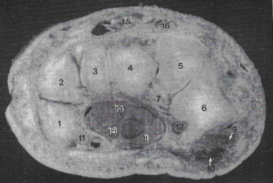 Figure 1. Photograph of transverse section of plastinated right human wrist. (1) pisiform, (2) triquetrum, (3) hamate, (4) capitate, (5) trapezoid, (6) trapezium, (7) scaphoid, (8) median nerve, (9) abductor pollicis longus muscle, (10) opponens pollicis muscle, (11) ulnar nerve, (12) flexor carpi radialis tendons, (13) flexor digitorum superficialis tendons, (14) flexor digitorum profundus tendons, (15) extensor digitorum tendons, (16) extensor carpi radialis. |
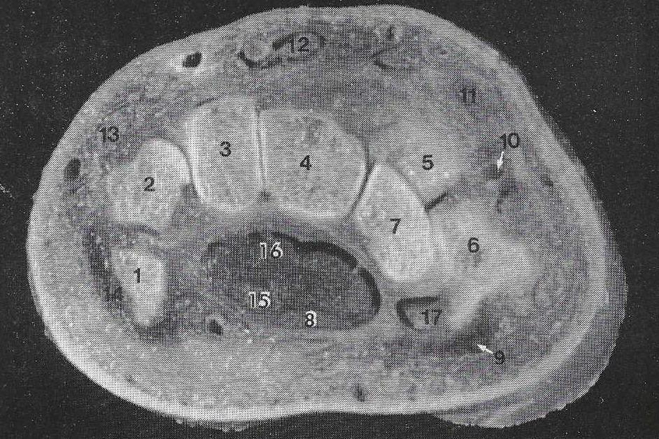 Figure 2. Transverse section of plastinated left human wrist. (1) pisiform, (2) triquetrum, (3) hamate, (4) capitate, (5) trapezoid, (6) trapezium, (7) scaphoid, (8) median nerve, (9) abductor pollicis brevis, (10) radial artery, (11) extensor pollicis longus tendon, (12) extensor digitorum tendons, (13) extensor carpi ulnaris, (14) digiti minimi muscles, (15) flexor digitorum superficialis tendons, (16) flexor digitorum profundus tendons, (17) flexor carpi radialis. |
Dehydration
The specimens were dehydrated in cold (-25 °C) acetone baths. In general, at least three changes of acetone were necessary, with each bath containing a volume of acetone 5-10 times the volume of the specimens. After dehydration was complete, the specimens were kept in room temperature acetone for one week for defatting.
Impregnation
After the specimens were defatted, the acetone served as an intermediary solvent and the specimens were immersed in the silicone resin, Biodur S 10 with S 3 Catalyst, in a vacuum chamber. Vacuum was applied and increased slowly over a four week interval. Impregnation was determined to be complete when acetone bubbles were no longer released from the specimen.
Curing
Excess resin was drained from the specimens and they were placed in a closed receptacle containing the curing agent S 6. A period of 4-6 days was required for the silicone polymer to completely harden. After which, the specimens were removed and ready for use.
The advantages of using plastinated specimens for teaching both anatomy and pathology is well known (Ostrom, 1987; von Hagens, 1979a; 1979b; 1985; 1988; Bickley, 1980; Bickley et al., 1981; Tiedemann and von Hagens, 1982. The process of plastination offers new alternatives for the study of anatomy. Gross specimens, dissected specimens, and cross-sectional slices can be preserved permanently into specimens which are clean, dry and practical to use, without the irritation and potentially harmful effects of older preservative liquids such as formaldehyde.
Anatomically, color and structural integrity of these specimens are far superior to other methods of preservation. This, along with their practicality, allows for several potential uses in academic and clinical medicine. Relationships of the flexor tendons and the median nerve, within the carpal tunnel, can be examined. As well as, identification of different tissue layers, nervous and vascular structures which lie within the osseous and ligamentous boundaries of the carpal tunnel.
Cross-sectional views can be studied in conjunction with either computer tomography or magnetic resonance imaging views of the wrist and hand. Other unexplored uses of such wrist specimens could be the practice of arthroscopic techniques in a distended plastinated specimen as is now being done with the knee (Tiedemann, 1988). As the use of plastination gains recognition, plastination will serve as a popular adjuvant to the teaching of orthopedics.
Baptista CAC, EP Cerqueira, JT Silva, JK Mansfield: Conservation and dry storage of cadavers through the vacuum for anatomical studies. Rev Bras Cienc Morf 3(2):121-123, 1986.
Bickley HC: Preservation of gross tissue specimens by plastination. Bull Pathol Educ 6:5-7, 1980.
Bickley HC, G von Hagens, FM Townsend: An improved method for preservation of teaching specimens. Arch Pathol Lab Med 105:674-76, 1981.
Oostrom K: Fixation of tissue for plastination: general principles. J Int Soc Plastination 1:3-11, 1987.
https://doi.org/10.56507/WLZH2223
Tiedemann K, G von Hagens: The technique of heart plastination. Anat Rec 204:295-299, 1982.
https://doi.org/10.1002/ar.1092040315
Tiedemann K: A silicone-impregnated knee joint as a natural model for arthroscopy. J Int Soc Plastination 2(1):13-17, 1988. https://doi.org/10.56507/CACT1479
von Hagens G: Impregnation of soft biological specimens with thermosetting resins and elastomers. Anat Rec 194:247-256, 1979a. https://doi.org/10.1002/ar.1091940206
von Hagens G: Emulsifying resins for plastination. Der Praparator 25:43-50,1979b.
von Hagens G: Heidelberg Plastination Folder: Anatomisches Institut 1, Universitat Heidelberg, 1985.
von Hagens G: Aldehyde-free embalming: some new developments. Fourth International Conference on Plastination, Mercer University School of Medicine, Macon, GA, 1988.