Departments of Anatomical Sciences1 and Pathology2 University of Oklahoma Health Sciences Center, College of Medicine P.O. Box 26901, Oklahoma City, OK 73190
A method for whole animal acetylcholinesterase (AChE) histochemistry was combined with standard S10 plastination. Resultant whole or partial animal specimens prepared in this manner were superior to whole animal specimens processed for AChE histochemistry and stored in formalin. The results compared favorably with those attained with isolated organ or tissue whole mounts while preserving and enhancing existing anatomical relationships. The technique was found to be useful in studying peripheral autonomic innervation by AChE histochemistry, in teaching specific anatomic details to laboratory personnel, and was suggested to be of potential use with other histochemical or immunonistochemical techniques in a variety of small animals, organs, or tissues.
AChe; S10; Plastination; Immunohistochemistry
Daniel L Feeback Departments of Anatomical Sciences University of Oklahoma Health Sciences Center, College of Medicine P.O. Box 26901, Oklahoma City, OK 73190

![]()


A variety of methodologic innovations have been devised to examine the distribution of autonomic nerves in organs and tissues of human and laboratory animals. In order to investigate patterns and properties of peripheral autonomic innervation, diverse neuro- histochemical techniques including enzymatic and immunohistochemical methods (Koelle and Friedenwald, 1949; Costa et al., 1980) have been applied to whole mounts of organs or organ laminae (Baljet and Drukker, 1975; Costa et al., 1980; Papka et al., 1981; Papka et al., 1985) or to paraffin or cryostat sections of organs (El- Badawi and Schenk, 1967; Papka et al., 1985; Schultzberg et al., 1979). Organ or tissue whole mounts are particularly useful for in situ demonstration of ganglia, ganglionated nerve plexuses, nerve trunks, and nerve fibers. Since anatomical relationships are preserved, these specimens are valuable for studying the density and distribution of nerve fibers and neuroeffector relationships.
We have developed a technique to produce permanent animal mounts by standard S10 plastination (Bickley et al., 1981; Bickley, 1980; von Hagens, 1979a; von Hagens et al., 1987; von Hagens, 1979b) of whole or party animal specimens following in situ histochemistry; in particular, localization of the enzyme acetylcholinesterase (AChE). Specimens prepared in this manner can be used as a reference during regional dissections of other animals to obtain tissue blocks for research purposes that contain specific anatomic structures of interest. They can also be utilized to teach laboratory personnel about important anatomic relationships as they relate to specific research projects. Plastinated whole or partial animal preparations are superior to organ or tissue whole mounts because additional anatomic relationships are maintained and they are permanent.
ANIMAL PERFUSION, SPECIMEN PREPARATION AND AChE
HISTOCHEMISTRY
An adult female Sprague-Dawley rat was anesthetized with intraperitoneal sodium pentobarbital (50 mg/kg) and fixed in toto by transcardial perfusion with normal saline (300 ml of 0.9% NaCl in water) followed by a fixative solution of 4% paraformaldehyde in 0.1 M phosphate buffer adjusted to a pH of 7.1. The total time of fixation was 4 hours. After fixation, regional abdominal and pelvic dissection was performed to expose the organs (intestinal tract, uterus, cervix, vagina) of interest to this laboratory. The urinary bladder and urethra were removed in order to reveal the ventral surfaces of the uterine cervix and vagina. Since we were primarily interested in the organs of the female genital tract, the body of the rat was transected at approximately the level of the kidneys and the rostral portion discarded. The caudal portion was washed thoroughly with 0.2 M acetate buffer (pH 5.5),and then preincubated in acetate buffer containing 10 M ISO- OMPA to inhibit nonspecific cholinesterase activity. The caudal body segment was incubated at ambient temperature and lighting for 16 hours in a medium containing acetylthiocholine iodide as substrate and 10" M ISO-OMPA. The medium was prepared In the following manner: acetylthiocholine iodide (500 mg) was dissolved in 20 ml of distilled water, and then 35 ml of a 1.0 M copper sulfate solution was added. The resultant solution was agitated thoroughly and centrifuged at 2000 rpm for 20 minutes. To 50 ml of dear supernatant, 310 mg of glycine was added, the pH was adjusted to 5.5 with 1.0 M sodium acetate, and the total solution volume was increased to 250 ml with distilled water. Next, the reaction product was developed by placing the specimen in a solution of ammonium sulfide [1 part ammonium sulfide (light) :22 parts distilled water] for one minute. The specimen was washed thoroughly in 0.2 M acetate buffer (pH 5.5), drained, blotted dry, and frozen in an ultracold freezer (-70 °C) for six hours pending plastination.
To compare the histochemical reaction product localization for AChE of this technique to that of isolated whole mount preparations of organs/structures, specific tissues were prepared as whole mounts. Rats were killed by an overdose of sodium pentobarbital (50 mg/kg, i.p.) followed by exsanguination. Fresh tissues (i.e. intestine and uterine cervix/vagina) were dissected in phosphate-buffered saline (PBS). The wall of the intestine was opened, gently stretched and pinned to balsa wood and fixed for 14-16 h in cold picric acid-2% formaldehyde in 0.1 M phosphate buffer, pH 7.1, whereas, the cervix/vagina preparation was fixed intact. These specimens were dehydrated in ethanol, cleared in xylene, and rehydrated in PBS. At this point, the different layers of the intestinal wall were separated into the outer longitudinal smooth muscle, the inner circular smooth muscle, the submucosa, and the mucosa. In these intestinal preparations, the myenteric plexus of nerves remains adherent to the outer muscle layer which produces a thin preparation facilitating the observation of nerves and ganglia. The cervix/vagina preparation required only the removal of excess fatty tissue. Acetylcholinesterase histochemistry was performed on both whole mount preparations (intestine and cervix/vagina) as described above.
S10 PLASTINATION
The caudal segment of the rat was dehydrated via freeze-substitution in -25 "C acetone for 6 weeks using 3 changes of acetone at 3, 2, and 1 week intervals. The specimen was submerged in a standard mixture of Biodur S10/S3 (von Hagens, 1985). After a 24 hour period of ambient pressure, vacuum was applied initially at 160 Torr and decreased by approximately 3 to 5 Torr daily over the next 4 weeks until the pressure reached 10 Torr. Subsequently, the pressure was decreased by 1 Torr each day until the pressure reached 4 Torr. At this time the system was completely closed with no additional air ingress. This setting was maintained for an additional 5 days until all bubble activity at the specimen surface had ceased.
Following forced impregnation and restoration of ambient pressure, excess polymer was drained from the specimen at -25 °C. For a period of 24 days, the specimen was removed daily from the freezer and placed in a closed gas cure chamber at room temperature. The chamber contained volatilized S6 gas and the specimen was exposed to this gas for increasing intervals. For the first 5 days, the specimen was exposed to 86 for 15 minute intervals (6 to 10 times per day) and then returned to the freezer for at least 30 minutes between gas exposure periods. Excess polymer was wiped from the surfaces of the specimen at least once during the cure period and always before returning the specimen to the freezer. On successive days, the length of exposure time was increased by approximately 5-10 minutes each day while the number of exposure periods per day was decreased. Finally the specimen was allowed to remain continuously in the gas cure chamber for 5 additional full days. The plastinated specimen was photographed with a Wild photomicroscope; whereas, photographs of the tissue whole mounts were taken on a Leitz Orthomat microscope.
The normal anatomic relationships of structures within the lower abdominal and pelvic cavities of the rat were maintained following AChE histochemistry and S10 plastination (Fig. 1). With the aid of a dissecting microscope, the dark histochemical reaction product resulting from the AChE activity of nerve fibers, ganglia, ganglionated nerve plexuses, and skeletal muscle motor end plates was readily visualized in the plastinated specimen (Figs. 2, 4, 5, 7). Selected areas from the plastinated specimen (Figs. 2, 5) were comparable in appearance and patterns of enzyme localization to corresponding areas from isolated whole mounts (Figs. 3, 6, respectively).
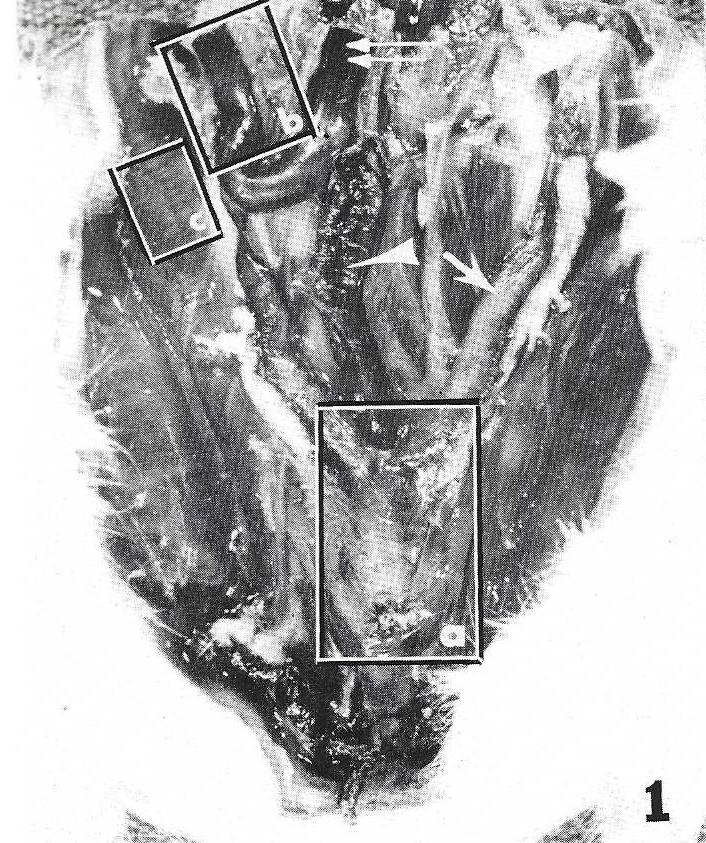 Figure 1. Low power macrophotograph of the pelvic and lower abdominal contents of a female rat processed in situ for AChE histochemistry. Caudal is toward the bottom of the photograph. Major landmarks include the colon (arrowhead), uterine horn (arrow), and small intestine (double arrow) . Box a outlines the uterine cervix and rostral vagina and is enlarged in Fig. 2. Box b outlines part of the gastrointestinal tract and is enlarged in Fig. 4. Box c outlines part of the lateral body wall and is enlarged in Fig. 7. |
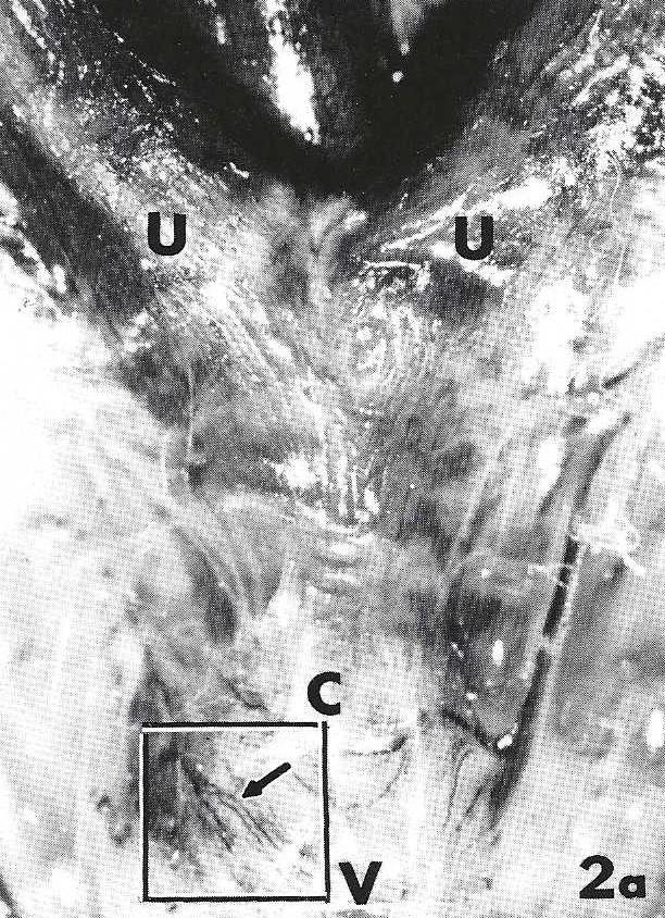 Figure 2a. Enlargement of box a of Fig. 1. AChE-positive nerves (arrow) are evident as they emerge from the paracervical ganglion and course across the cervix-vagina junction. Uterine horns (U), Cervix (C), Vagina M. X 8.6. |
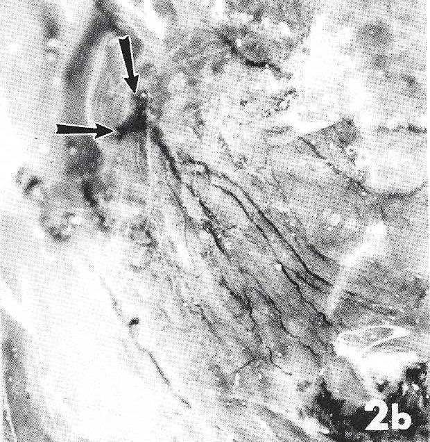 Figure 2b. Enlargement of the area in the box of Fig. 2a. Note the AChE-positive nerves emanating from the paracervical ganglion (arrows). X 13.5. |
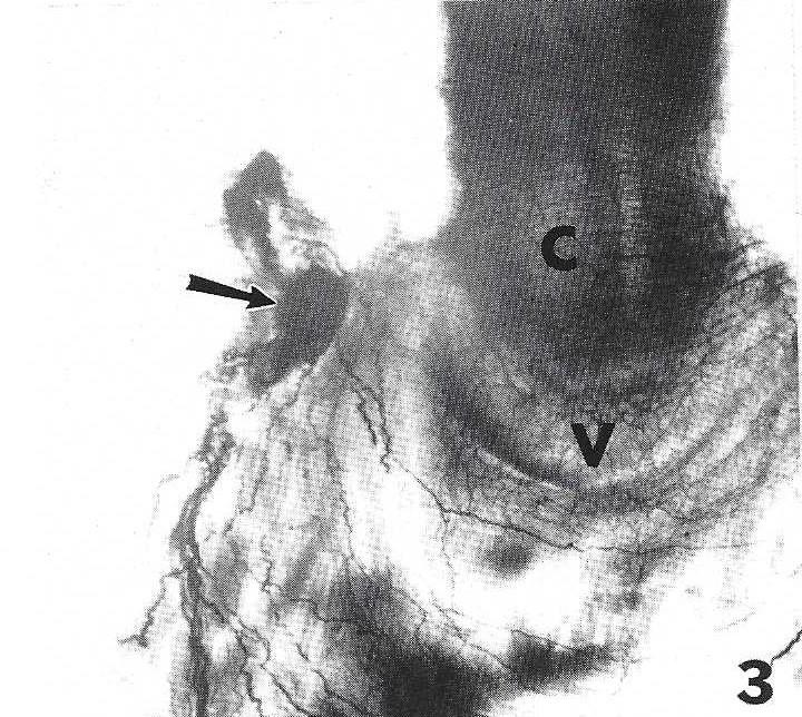 Figure 3. Isolated whole-mount preparation of the uterine cervix (C) - vagina M junction comparable to the area illustrated in Fig. 2. AChE-positive nerves stem from the paracervical ganglion (arrow) and form plexuses associated with the cervix and vagina similar in pattern to those observed in whole body preparations. X 13.5. |
Low power microscopy of the uterus, cervix, and vagina (Figs. 1, 2a) demonstrated the presence of AChE- positive nerve fibers. Their density and distribution as they radiated out from the paracervical ganglion toward the junction of the cervix with the vagina was evident (Figs. 2a, 2b).
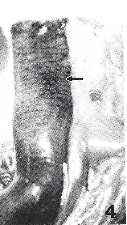 Figure 4. Low magnification of intestine (box b, Fig. 1). Even though some parts of the gut are slightly out-of-focus, it is possible to appreciate the arrangement and density of the AChE-reactive myenteric plexus (arrow) around the wall. X 6.8. |
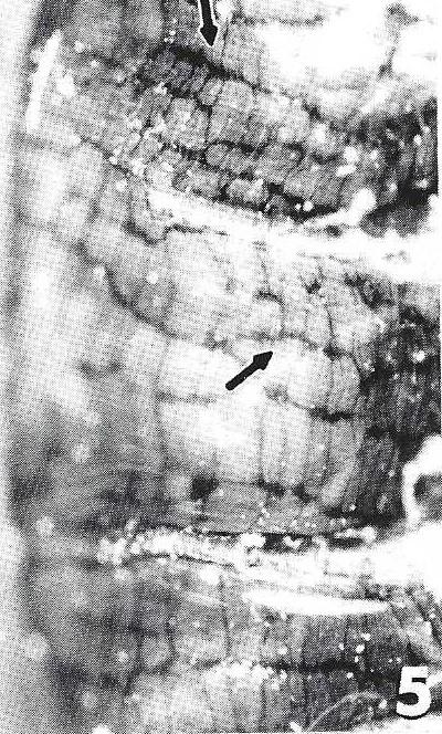 Figure 5. Higher magnification of the intestinal wall showing the arrangement of the AChE-positive nerves forming the myenteric plexus (arrow) . X 9.2. |
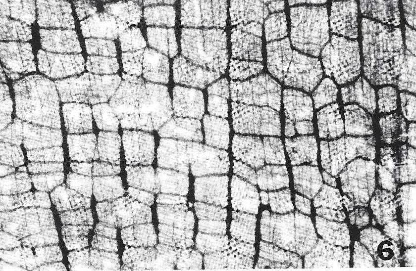 Figure 6. Whole-mount preparation of the myenteric plexus of the gut, histochemically stained, demonstrating the AChE-positive ganglionated nerve plexus. X 10.8. |
|
In intestine (Fig. 4) at low magnification, the expected AChE-reactivity of the ganglionated myenteric plexus (Fig. 5) was observed. The net-like appearance of the plexus was similar to that achieved with AChE histochemistry of isolated whole mount preparations (Fig. 6). Motor end plates appeared as discrete structures aligned along the muscle motor point (Figs. 7a, 7b) in the skeletal muscle of the lateral body wall.
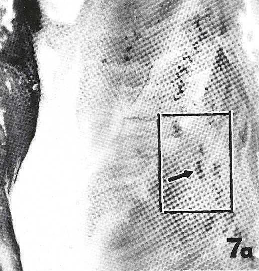 Figure 7a. Low magnification of the body wall musculature (box c, Fig. 1): Note numerous AChE-positive motor endplates (arrow) aligned at the muscle midpoints. X 9.2. |
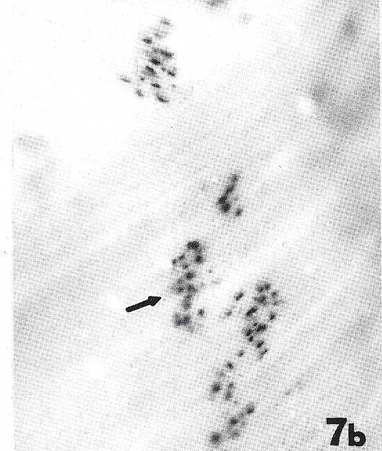 Figure 7b. Higher magnification (boxed area, Fig. 7a) demonstrating detail of the motor endplates. X 23.4. |
Whole animal preparations offer advantages over whole mounts of organs or organ laminae, since the latter are limited to examination of single organs or to portions of a single organ. Whole animal preparations demonstrate and preserve anatomic relationships that exist between organs and functionally related structures. A major problem with the current methods for preparing whole mounts is that most organs must be separated into laminae or physically disrupted in some manner to gain the resolution needed for microscopy. In addition, they must be stored in fixative (usually formalin) and therefore have a limited useful life. The disadvantages inherent to fixed specimens (unpleasant odor; irritation to eyes, skin and mucous membranes; necessity to maintain a moist environment; and discoloration and deterioration with prolonged storage) make this method of preservation and storage less than optimal.
Use of standard 810 technique to plastinate whole mounts stained for acetylcholinesterase (AChE), results in specimens superior to those stored in formalin that compare favorably with isolated organ or tissue whole mounts in which AChE activity has been demonstrated. Plastinated whole animal specimens are particularly valuable for demonstrating to laboratory personnel the proper tissue blocks to remove from experimental animals for preparing whole mounts of organs or organ laminae or for freezing and cryostat sectioning. We suggest that this method could be used for other types of histochemical, and possibly immunohistochemical procedures in a variety of small animals, organs, and tissues.
ACKNOWLEDGEMENTS:
Supported in part by NIH Grant NS22526, the Presbyterian Health Foundation, and a grant from the University of Oklahoma College of Medicine Alumni Association.
Baljet B, J Drukker: An acetylcholinesterase method for In toto staining of peripheral nerves. Stain Technol 50:31-36, 1975. https://doi.org/10.3109/10520297509117028
Bickley HC, G von Hagens, FM Townsend: An improved method for preserving specimens. Arch Pathd Lab Med 105:674-676, 1981.
Bickley HC: Preservation of gross specimens by plastination. Bull Pathol Educ 6:5-7,1980.
Costa M, R Buffa, JB Furness, E Solcia: Immunohistochemical localization of polypeptides in peripheral autonomic nerves using whole mount preparations. Histochemistry 65:157-165,1980.
https://doi.org/10.1007/BF00493164
El-Badawi A, EA Schenk: Histochemical methods for separate, consecutive and simultaneous demon- stration of acetylcholinesterase and norepinephrine in cryostat sections. J Histochem Cytochem 15:580-588, 1967.
https://doi.org/10.1177/15.10.580
Koelle GB, JS Friedenwald: A histochemical method for localization of cholinesterase activity. Proc Soc Exp Biol Med 70:617-622, 1949. https://doi.org/10.3181/00379727-70-17013
Papka RE, JP Cotton, HH Traurig: Comparative distribution of neuropeptide tyrosine-, vasoactive intestinal polypeptide-, substance P-immunoreactive, acetylcholinesterase-positive and noradrenergic nerves in the reproductive tract of the female rat. Cell Tissue Res 242:475-490,1985.
https://doi.org/10.1007/BF00225412
Papka RE, JB Furness, NG Delia, M Costa: Depletion by capsaicin of substance P-immunoreactivity and acetylcholinesterase activity from nerve fibres in the guinea-pig heart. Neurosci Lett 27:47-53,1981.
https://doi.org/10.1016/0304-3940(81)90204-4
Schultzberg M, T Hokfelt, L Terenius, L-G Elfvin, JM Lundberg, J Brandt, RP Elde, M Goldstein: Enkephalin immunoreactive nerve fibres and cell bodies in sympathetic ganglia of the guinea-pig and rat. Neuroscience 4:249-270,1979. https://doi.org/10.1016/0306-4522(79)90087-3
von Hagens G: Emulsifying resins for plastination. Praparat 24:43-50,1979a.
von Hagens G: Impregnation of soft biological specimens with thermosetting resins and elastomers. Anat Rec 194:247-256, 1979b. https://doi.org/10.1002/ar.1091940206
von Hagens G: Heidelberg Plastination Folder: Collection of all technical leaflets for plastination. Anatomisches Institut 1, Universitat Heidelberg, 1985.
von Hagens G, K Tiedemann, W Kritz: The current potential of plastination. Anat Embryol 175:411-421, 1987.
https://doi.org/10.1007/BF00309677