Departments of Biology1 & Biomedical Sciences2, Grand Valley State University
Allendale, Michigan, U.S.A.
Here we describe plastination of a bowfin fish (Amia calva) and post-plastination dissection of the plastinate conducted to expose the articulation between the skull and pectoral girdle and the individual elements of the pectoral girdle. The specimen produced during this project demonstrates that useful anatomical relationships can be revealed by dissection after the plastination process has been completed.
Amia; pectoral girdle; post-plastination dissection
Bruce D. Ostrow: Departments of Biology, Grand Valley State University, Allendale, Michigan, U.S.A. 49401. Telephone: 616-331-8581; Fax: 616-331-3446; E-mail: ostrowb@gvsu.edu.

![]()


The skull and pectoral girdle of the bowfin fish (Amia calva) are very useful teaching aids to demonstrate skull and forelimb evolution to comparative vertebrate anatomy students. Amia retains elements of the ancestral vertebrate skull and pectoral girdle that are easy to view relative to other extant teleosts. Skeletal material is readily available from biological supply firms (e.g. Ward’s Natural Science), but the skull is never supplied with the pectoral girdle attached, requiring the students to approximate the relationships of these two articulating elements. A much better appreciation of the two elements can be gained from an intact specimen that has been dissected to show the area of connection. The idea of such a dissection occurred after a 61cm formalin-preserved Amia which had been used to show external features to comparative anatomy students had been plastinated to facilitate its storage and use in the course. Out of necessity, we decided to dissect our only plastinate to expose the pectoral girdle. The results of the dissection are shown and the process described briefly here.
Specimen procurement
A 61cm bowfin fish (Amia calva) was captured in an unnamed freshwater lake in Michigan, (USA) and donated to our institution. It was fixed whole in 10% formalin buffered with disodium phosphate to prevent decalcification of bones. It had been stored for approximately 13 years in the same solution.
Protocol for plastination
We subjected the specimen to room-temperature plastination (Zheng et al., 1996; Raoof et al., 2007). In every step, a stainless steel grate was used to keep the specimen submerged.
To expose the body cavity of the fish to the plastination solutions, the left lateral flank of the fixed specimen was opened with a scalpel and a rectangular section of body wall was removed en bloc (Fig. 1). The fixed, dissected specimen was washed in cold running water for 2 days to remove excess formalin fixative.
The specimen was dehydrated initially in 10 specimen volumes of 90% acetone (Fisher Scientific) for 24 hr at room temperature, then in three sequential changes of 10 specimen volumes of 100% acetone at room temperature. Acetone concentration was checked with an ethanol hydrometer (Fisher Scientific 2510C) until it stabilized at >99%.
The dehydrated specimen was soaked in 10 L 93% Cortech PR-10 polymer (Dow Corning), 7% Cortech CR-22 crosslinker (Dow Corning) for 24 hr at room temperature, then vacuum was applied. To reduce the risk of shrinkage, tank pressure was decreased gradually in 6 inch increments to 29 inches Hg (736mm Hg) over the first 24 hours and maintained at that level for three days until bubbles no longer emanated from the specimen. The impregnation tank was brought to atmosphere over a two-hour time period by a bleed-valve. The specimen was removed immediately and drained of excess polymer/crosslinker head-down for
24 hours.
Curing was accomplished by spraying on Cortech CT-32 catalyst (Dow Corning) and wrapping in plastic wrap for 16 hours.
Post-plastination dissection
Because we had neglected to expose some relevant anatomical structures in the original dissection of our specimen, out of necessity the plastinate was further dissected. Skin and superficial muscle tissue was cut transversely using a scalpel and removed with forceps. Deeper muscles were scraped from the bones with the flat edge of the scalpel blade.
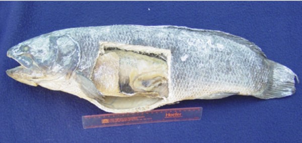 Figure 1 - Amia plastinate. Note the internal organs exposed by opening in flank and the lack of an opening between the gill operculum and pectoral fin. |
The initial dissection removed a section of the left lateral body wall exposing the internal abdominal organs. The remaining specimen was subjected to room temperature plastination (Fig. 1).
In order to expose the articulation between the skull and pectoral girdle, the plastinated specimen was further dissected to expose the region of articulation (Fig. 2). We also dissected to expose the individual elements of the pectoral girdle; specifically the posttemporal, supracleithrum, postcleithrum, cleithrum, and scapulocoracoid (Figs. 2 and 3). Terminology is that of Grande and Bemis (1998). The plastinated skin and associated scales were rather sturdy and presented the greatest challenge to slice through, but removal of deeper muscle and connective tissues was relatively easy. Deep muscles had the same texture as superficial muscles, indicating thorough impregnation of the plastination solutions and curing catalyst. Muscles retained structure and were cut out en bloc or scraped off the bones. Scraping of muscle tissue produced fluffed material which was easily removed. We found that dissection of individual plastinated muscles was difficult, primarily because the plastinated muscles were quite friable and not easily separated intact. We did a small-scale exploratory dissection of a plastinated shark (Squalus sp.) pectoral girdle and found that the muscle tissue was similar to that of the bowfin plastinate (data not shown).
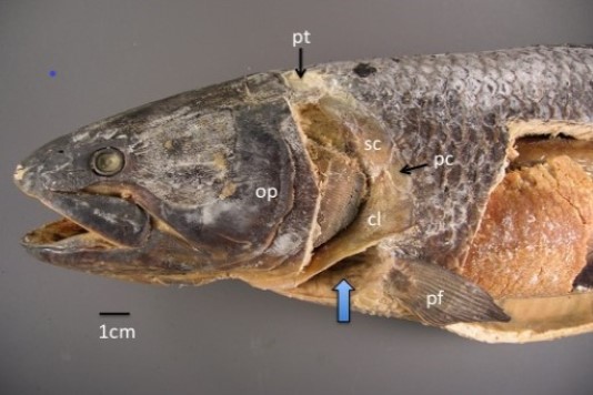 Figure 2 - Lateral view of anterior half of Amia plastinate after post-plastination dissection. Note the two new cavities relative to figure 1: one posterior to the gill operculum (op) and one anterior to the pectoral fin (pf, up arrow). The pectoral girdle elements that are visible are the posttemporal (pt), supracleithrum (sc), postcleithrum (pc), and cleithrum (cl). |
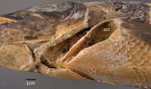 Figure 3 - Ventrolateral view of Amia plastinate after post-plastination dissection. The scapulocoracoid (scc) is visible in the posterior opening deep to the cleithrum (cl). pc = postcleithrum. |
The finished specimen was used as a teaching tool in a Comparative Vertebrate Anatomy course at our institution. All individual bones of the pectoral girdle were readily displayed, and the specimen now shows the skull and pectoral girdle in context to one another and to the rest of the specimen. Students were surveyed as to the usefulness of the plastinate for their understanding of the pectoral girdle relative to a partially articulated pectoral girdle in a display box. Sixteen out of 18 students reported that they preferred the plastinate. One student reported it was “helpful to see how it shows the relationship better.”
The core steps of plastination are fixation, dissection, dehydration, forced impregnation, and curing (von Hagens et al., 1987). To this sequence of procedures we add an optional step: dissection after curing. We found that this plastinated fish specimen had sufficient structure to support the exposed skeletal structure and to withstand dissection, and had sufficient malleability to allow dissection with traditional dissection instruments.
Opening the flank exposed the internal organs to the plastination solutions. Perhaps we could have plastinated the specimen whole but we deemed it prudent to perforate the flank to increase the likelihood of solution penetration through the thick, tough body wall.
We chose room temperature dehydration and plastination because we were not concerned about shrinkage with this specimen due to lack of fatty tissue (Raoof et al., 2007). If one were to attempt this procedure with a fatty specimen or neural tissue, we recommend cold-temperature (-20°C) plastination to reduce shrinkage.
The plastinate’s body wall was rather sturdy and presented the greatest challenge to dissection, but removal of deeper muscular and connective tissues was relatively easy. Scraping of deeper muscle tissue produced fluffed material which was easily removed. However, it appeared that post-plastination muscle dissection to identify individual muscles is difficult because the plastinated muscles are quite friable and not easily separated intact, as is the case with fresh or formalin-preserved material. Prior to plastination, formalin-fixed muscle tissue has the texture of a rubber bicycle tire. We found that after plastination, muscle tissue is stiff and friable.
Plastination of fish has been accomplished (Asadi, 1998), but to our knowledge, whole and not dissected after plastination.
The specimen produced during this project was well-preserved and suitable for use as a teaching tool. Although dissection of fresh or formalin-preserved specimens may give superior results, it is noteworthy that useful material can also be produced by dissection after the plastination process has been completed, even years post-plastination. Once cured, the plastinate is stable, intact, and can be used or dissected at any later date. We happened to wait over one year to do the post-plastination dissection.
Acknowledgements
We thank Dr. Ameed Raoof of the University of Michigan for allowing us access to the Plastination Laboratory. We thank also anonymous reviewers for constructive criticism.
Asadi, MH. 1998: Plastination of sturgeons with the S10 technique in Iran: the first trials. J. Int. Soc. Plastination 13:15-16.
https://doi.org/10.56507/XSTD4829
Grande L, Bemis WE. 1998: A comprehensive phylogenetic study of amiid fishes (Amiidae) based on comparative skeletal anatomy. An empirical search for interconnected patterns of natural history. Society of Vertebrate Paleontology Memoir 4: i-x, 1-690; supplement to Journal of Vertebrate Paleontology 18(1).
https://doi.org/10.1080/02724634.1998.10011114
Raoof A, Henry RW, Reed RB. 2007: Silicone plastination of biological tissue: room-temperature technique DowTM/Corcoran technique and products. J. Int. Soc. Plastination 22:21-25.
https://doi.org/10.56507/AWAC9285
von Hagens G, Tiedemann K, Kriz W. 1987: The current potential of plastination. Anat Embryol 175: 411-421.
https://doi.org/10.1007/BF00309677
Zheng TZI, Weatherhead BI, Gosling J. 1996: Plastination at room temperature. J. Int. Soc. Plastination 11:33.
https://doi.org/10.56507/EHWO1033