Plastination is a process of long-term preservation of biological tissue. This process is gaining popularity for its benefits as a teaching and research tool in anatomy. The process is based on replacement of water and fat by forced impregnation, after replacing the water by an intermediate solvent, to produce hard, dry and odourless specimens. However, due to the specialized equipment and expensive chemicals, such as polymer, that are needed for plastination, we desired to simplify the process and embed the samples completely in paraffin wax. In this process, the water and fat were replaced by paraffin, yielding specimens that can be touched, that do not smell or decay, and that even retain most properties of the original sample. Various freshly-collected visceral organs of goat were used for preparing specimens. These organs were dissected out to expose different gross anatomical features. The specimens were fixed in 10% formalin, hardened in a deep freeze, dehydrated and dried in freeze dryer, impregnated with melted paraffin, cured, and then stored at room temperature for further use as educational tool. The prepared wax-impregnated specimens were clean, dry, odorless, durable, non-toxic, and can be handled by bare hands and do not require any special storage care. This method will strengthen the appearance, clarity of surface anatomy, and description of other parts, as well as practical demonstrations in undergraduate teaching, and enhance the anatomical museum collection. The anatomical accuracy and durability of these specimens make them powerful tools to accelerate knowledge acquisition, and strengthens diagnostic abilities for veterinary students utilizing a wider variety of learning strategies.
goat; internal organs; paraffin; plastination
Dr. Nasrin Sultana, Associate Professor, Department of Anatomy and Histology, Faculty of Veterinary Science, Bangladesh Agricultural University, Mymensingh 2202, Bangladesh, Email: nsultana.bau@gmail.com

![]()


The animal cadaver and its organs are an integral part of learning veterinary anatomy in the DVM program. The specimens used in teaching may be fresh or preserved (Ameko et al., 2012). However, decay of fresh specimens in a short time is a serious problem, so the specimen must be preserved to remain intact for prolonged periods by traditional methods (Mohamed and John, 2018). Even today, the most common practice is that anatomical, pathological, and other biological specimens are preserved and stored in 10% formalin solution or in Keyserling solution for the purpose of display and future study (Fischer, 1905; Siddiqui et al.,1988; Kumar et al., 2005).
These solutions are toxic, allergenic, and possibly carcinogenic. Concentrations of formaldehyde above 0.1 ppm in air can irritate mucous membranes and cause watery eyes. Moreover, inhalation of formaldehyde at the same concentration may cause breathing difficulties, a burning sensation in the throat, headache, and may even lead to asthma. It is troublesome to handle the formalin-fixed animals for teaching and research purposes, and, after a long period of time the quality of the specimens can also deteriorate (de Jong and Henry, 2008). To overcome these problems, Dr. Gunther von Hagens, introduced a new technique, plastination, to the medical world (von Hagens, 1985, 1986).
Plastination is a process for the long-term preservation of biological tissues, with a wide variety of processes and developments. The process is based on the idea of replacing the tissue water by an intermediate solvent that is then replaced by the polymer, by forced impregnation, to produce hard, dry and odorless specimens (Weiglein,1997). In recent years, there has been a growing trend toward plastinated products (Jones and Whitaker, 2009). Plastination provides a highly useful method for study that is gaining increased popularity for its utility in anatomy teaching and research (Ravikumar, 2014).
However, due to the specialized equipment and expensive chemicals, such as polymer, that are needed for plastination, we desired to simplify the process and embed the samples completely in paraffin. In this process, the water and fat are replaced by paraffin, yielding specimens that can be touched, do not smell or decay, and even retain most properties of the original sample. The process described here is a promising and economical method for preserving specimens that is a viable alternative to formalin preservation (Dawson et al., 1990).
Experimental animals were collected from the local market. The animals were euthanized for the prevention of cruelty to animals. The fresh organs were removed from the euthanized goats for further processing.
Specimen preparation
For dissecting the animals, a median incision was made from the mandibular space (between the rami of the mandible) to the anus. The body trunks were opened to show the internal organs in situ. Then, the organs of the digestive system, respiratory system, and urogenital tract, as well as the heart, spleen, and brain were collected. The tubular and hollow organs were washed for 3-5 minutes and packed with cotton-gauze to preserve their proper shape.
Fixation
Formalin is a commonly-used fixative in embalming fluid to avert decomposition, and deactivate saprophytic bacteria, thus preventing putrefaction. For fixation, collected organs from different systems of the body were preserved in 10% formalin for one (1) week to ensure proper fixation (Fig. 1).
Hardening
The fixed samples were washed in running tap water overnight and then placed in a deep freeze until a smoky layer appeared on the surface of the specimens (Fig. 2).
Dehydration and drying
All the specimens prepared for paraffin-embedding were placed in the vacuum chamber of a freeze dryer (Fig. 3). The freeze dryer removes all ice and frozen solvents from the specimens through the process of sublimation. It also removes bound water molecules through the process of desorption. In the end, the specimens become light in weight.
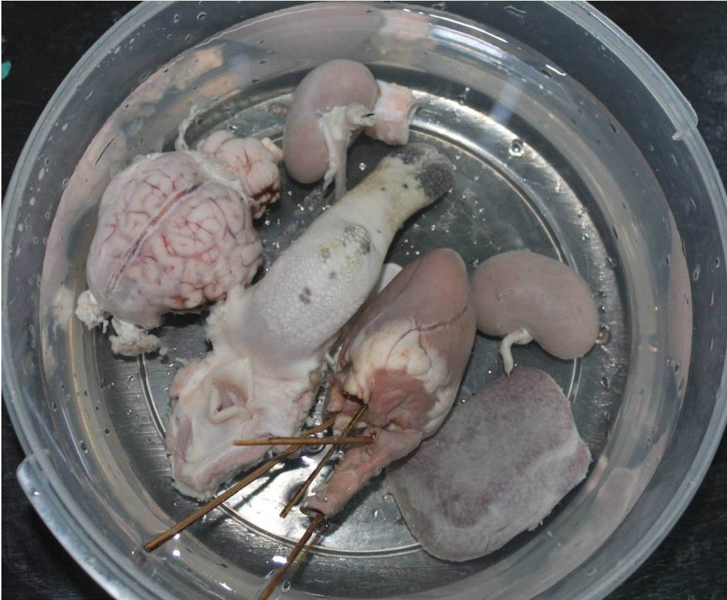 Figure 1. Representative samples (brain, tongue, heart, kidney and spleen of goat) during the fixation process in 10% formalin. |
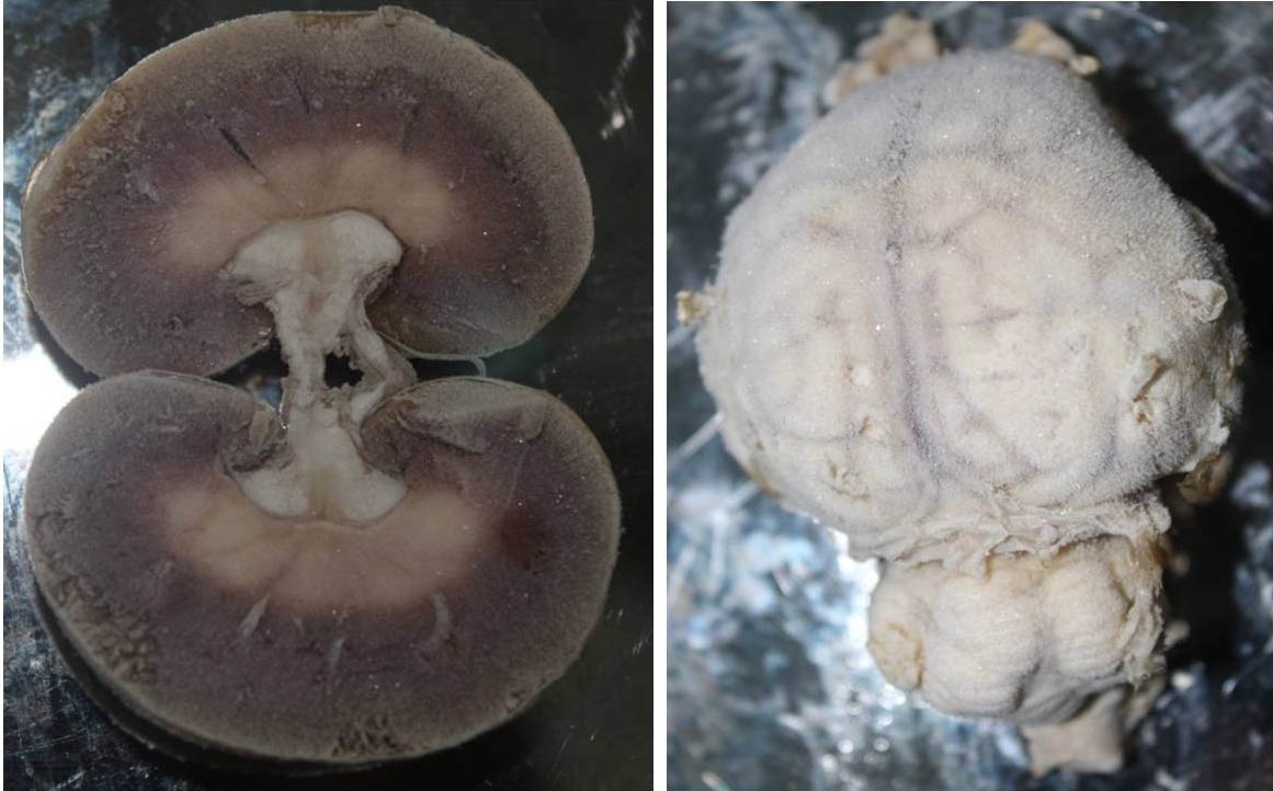 Figure 2. Representative samples (kidney and brain of goat) of hardening process with the appearance of smoky layers in the samples after removal from deep freeze (-20⁰). |
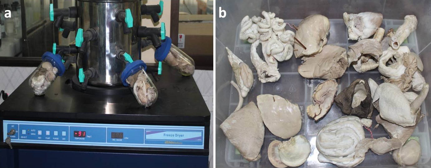 Figure 3. Samples drying in a freeze dryer (IlshinBioBase) (a), and the samples after freeze drying (b). |
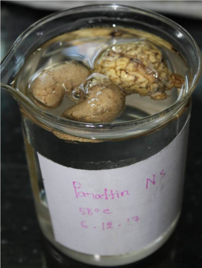 Figure 4. Impregnation of samples in melted paraffin (58° C). |
Impregnation
Impregnation of the specimens was performed in a chamber containing melted paraffin (Fig. 4). For impregnation, the dehydrated specimens were immersed in melted paraffin in an oven. After two dips in melted paraffin (each dip consisted of 1 minute in 60⁰ C melted paraffin), the specimens were kept at room temperature for hardening. Once the impregnation step was completed, the specimens were placed in a closed container containing silica gel desiccant.
Finishing and storage
The impregnated specimens were trimmed of excess unwanted solid paraffin from the edges of organs using a scalpel.. Finally, the samples were stored in a plastic bag at room temperature.
The internal or visceral organs (those of the digestive, respiratory, urinary, genital, cardiovascular, and nervous systems) of goat were impregnated by paraffin for use in practical anatomy demonstrations. After impregnation, the internal organs from the thoracic, abdominal, and pelvic cavities retained their original shape. These impregnated specimens (Figs. 5, 6, and 7) did not lose any aspects of their physical appearance, such as color, following the impregnation process. Their surfaces were dry and were able to be handled using bare hands. The prepared specimens could then be easily stored for a longer period of time without any preservatives.
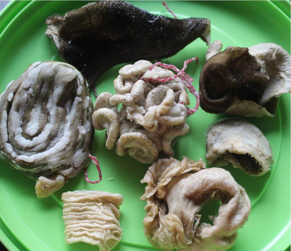 Figure 5. Representative paraffin-impregnated specimens of the digestive system (rumen, reticulum, omasum, duodenum, cecum, colon and rectum) of goat. |
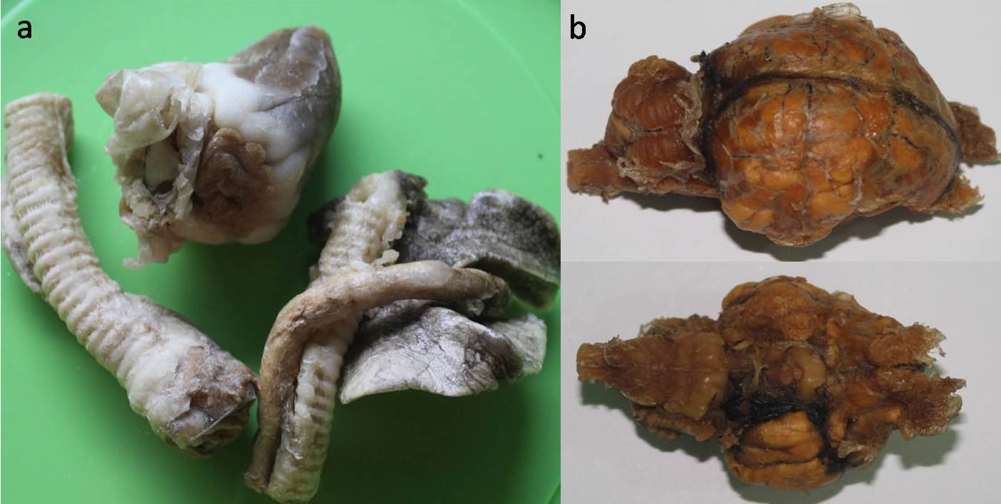 Figure 6. Representative paraffin-impregnated specimens of the respiratory system (trachea and part of lung) (a), and nervous system (dorsal and ventral view of brain) (b) of goat. |
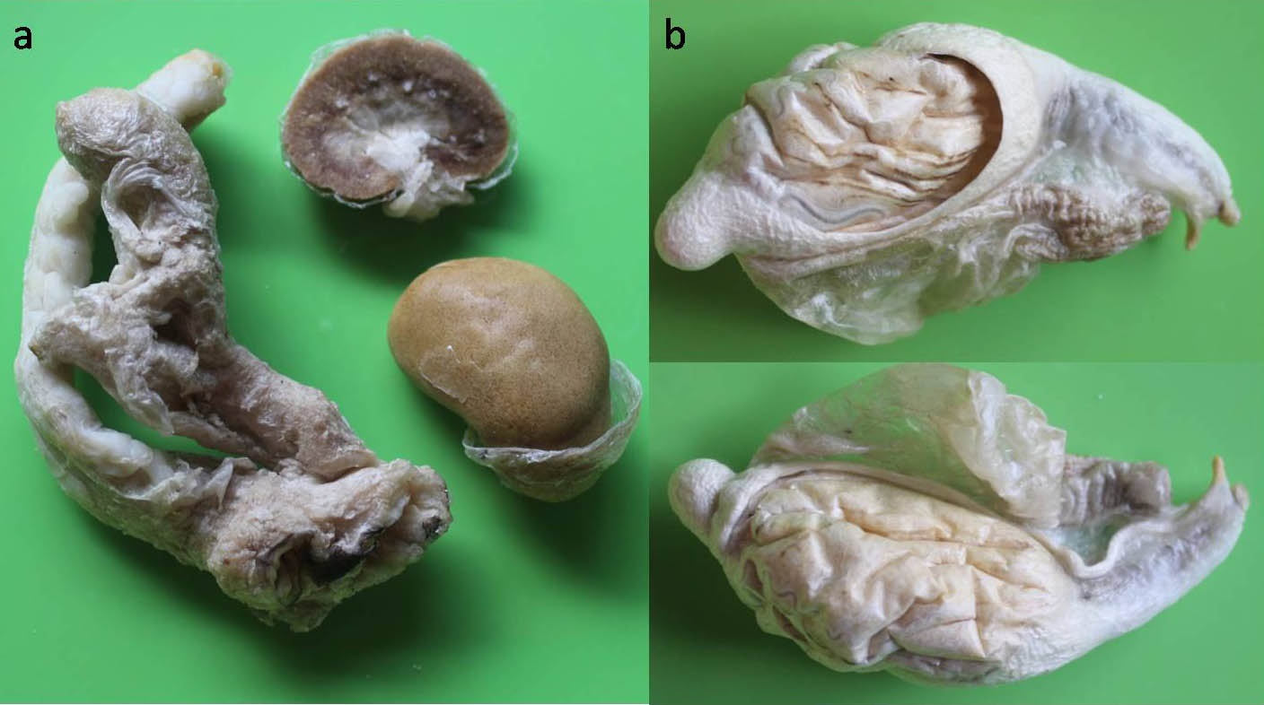 Figure 7. Representative paraffin-impregnated specimens of the urinary system (kidney, ureter and urinary bladder) (a), and genital system (testis with scrotum and epididymis) (b), of goat. |
Importance of the plastination technique
The applications and advantages of the plastination technique have been widely reported in the fields of training, research, and education (Chandini, 2014). Its major advantages include the plastinates being dry, free of offensive odor, durable, and safely handled using bare hands (Timothy et al., 1990; Pashaei, 2010; Chaturvedi et al., 2014). These specimens do not appear artificial in any way and do not require any special care or conditions for storage. The importance of the plastination technique has been well described in anatomical education (de la Torre et al., 2004), being reported as a basic method of anatomical investigation. Plastination facilitates contact by anatomy students, and also reduces exposure to irritants, such as formalin, through inhalation during practicals. In anatomy practical classes, the samples used for teaching students are either fresh or preserved. When using fresh samples, it is necessary to buy a new animal each time, while on the other hand, unnecessary storage in formalin can pose health hazards. For a complete academic year, a number of animals are necessary for dissection purposes alongside formalin-preserved samples. Therefore, plastination is an economical alternative to the traditional method of anatomical sample preservation when teaching anatomy. In a study of an undergraduate anatomy course at Cambridge University, plastinated specimens were used alongside wet cadaver dissection in anatomy practical lessons (Latorre et al., 2016). The plastinated specimens occupied less space in the anatomy laboratory than the formalin-preserved ones which required containers. Also, the formalin needed to be changed regularly to prevent mold growth. Similarly, the plastination technique has commonly been used not only for anatomical specimens but also for pathological specimens in undergraduate education (Bickley et al., 1987(b); Ravi and Bhat, 2011).
Principles of plastination techniques
The three recognized methods of plastination are silicone plastination, sheet plastination with epoxy, and sheet plastination with polyester (Sargon and Tatar, 2014). Of these three techniques, silicone plastination is the most common, in which fresh or formalin-fixed samples can be plastinated. This method can be used for cadavers, organs, and tissue slices. Epoxy plastination is done only for sliced (2-5 mm thickness) biological specimens. Epoxy and polyester plastination utilize the same basic principles as silicone plastination, with four steps: fixation, dehydration and defatting, forced impregnation, and curing (Bickley et al., 1987(a)). For fixation, formalin solution is used in a concentration between 5% and 20%. Cryosubstitution is the method of choice for dehydration, in a series of -20° C acetone baths. Forced impregnation is performed in a vacuum chamber; the acetone-filled specimens are submerged in a bath of polymer, where the acetone in the specimen is replaced with the polymer of choice using the force of the increasing vacuum. After removal from the polymer bath, the specimens are placed in an airtight container. Initially, polymer oozes out from the surface of the specimens. After a few days, the specimens can be finally stored in a airtight container containing desiccant, after curing by gas or catalyst.. The technique described here is a modification of the silicone plastination technique, to facilitate ease of use and economy. Dehydration is performed in a freeze dryer followed by drying. The impregnation step is performed in melted paraffin in an oven. Finally, the samples are stored in polybags with desiccant in a container.
Acknowledgements:
The authors acknowledge the expert technical assistance of the Anatomy Departments of Bangladesh Agricultural University and Bangladesh Institute of Nuclear Agency, Mymensingh, Bangladesh. We would like to thank Dr. Nabiul Islam, Assistant Professor of the Department of neuroscience at Yamaguchi University Graduate School of Medicine for his critical reading of the manuscript. We would also like to thank Dr. Greggory Wroblewski of the Center for Language Education at Ritsumeikan Asia Pacific University in Japan for his proofreading of the manuscript. Dr. Nasrin Sultana received research funds from the Ministry of Science and Technology, Bangladesh.
Ameko E, Achio S, Alhassan S, Adasu C, Dzagbletey E, Abbey PR. 2012: Plastination of some cow and ram organs in Ghana for use as teaching aids. Int J Pure Appl Sci Technol. 8: 57-68.
Bickley H C, Robert S C, Anna NW, Robert LJ. 1987a: Preservation of tissue by silicone rubber impregnation. J Int Soc Plastination 1: 30-39.
https://doi.org/10.56507/XVDP9663
Bickley HC, Walker AN, Jackson RL, and Donner RS. 1987b: Preservation of pathology specimens by silicone plastination. An innovative adjunct to pathology education. Am J Clin Pathol. 88: 220-223.
https://doi.org/10.1093/ajcp/88.2.220
Chandini R. 2014: Plastination. J. Pharm. Sci. & Res. 6: 271-273.
Chaturvedi RK, Anurag S, Praveen C, Abha M, Mishra SP. 2014: Advantages of Plastinated Human Body in Medical Education and its Legal & Ethical Aspects. J of Evolution of Med and Dent Sci. 3: 2626-2631.
https://doi.org/10.14260/jemds/2014/2184
Dawson TP, James RS, and Williams GT. 1990: Silicone plastinated pathology specimens and their teaching potential. J Pathol. 162: 265-272.
https://doi.org/10.1002/path.1711620314
de Jong K, and Henry RW. 2007: Silicone plastination of biological tissue: Cold-temperature technique BioDur S10/S15 technique and products. J Int Soc Plastination. 22:2-14.
https://doi.org/10.56507/ZLMJ7068
Fischer MH. 1905. Toxic effects of formaldehyde and formalin. J Exp Med. VI: 487-518.
https://doi.org/10.1084/jem.6.4-6.487
Jones DG and Whitaker MI. 2009: Engaging with plastination and the Body Worlds phenomenon: A cultural and intellectual challenge for anatomists. Clin. Anat., 22:770-6.
https://doi.org/10.1002/ca.20824
Kumar R, Nagar JK and Gaur SN. 2005. Indoor air pollutant and respiratory morbidity. Indian J Allergy Asthma Immunol. 19: 1-9.
Latorre R, Bainbridge D, Tavemore A, and LopezAlbors O. 2016: Plastination in Anatomy Learning: An Experience at Cambridge Universit. J Vet Med Educ. 43: 226-34.
https://doi.org/10.3138/jvme.0715-113R1
Mohamed R and John R. 2018: Production and use of plastinated anatomical specimens as teaching and learning tools in veterinary gross anatomy in the Caribbean. J Adv Vet Anim Res. 5: 44-52.
https://doi.org/10.5455/javar.2018.e245
Pashaei S. 2010: A brief review on the history, methods and applications of plastination. Int J Morphol. 28: 1075-1079.
https://doi.org/10.4067/S0717-95022010000400014
Ravi SB and Bhat VM. 2011: Plastination: A novel, innovative teaching adjunct in oral pathology. J Oral MaxillofacPathol. 15: 133.
https://doi.org/10.4103/0973-029X.84475
Ravikumar C. 2014: Plastination. J.Pharm. Sci. & Res. 6: 271-273.
Sargon MF and Tatar l. 2014: Plastination: basic principles and methodology. J Exp Clin Anat. 8:13-18.
https://doi.org/10.2399/ana.14.040
Siddiqui MA, Akbar M, Rana MW. 1988: Plastination technique for Museum specimens. JAMC. 3: 23-26.
Timothy PD, James RS, Williams GT. 1990: Silicone plastinated pathology specimens and their teaching potential. J pathol. 162: 265-272.
https://doi.org/10.1002/path.1711620314
de la Torre FR, Rodríguez-Baeza A, and Doménech-Mateu JM.2004: Setting up a plastination laboratory at the Faculty of Medicine of the Autonomous University of Barcelona. Eur J Anat, 8: 1-6.
von Hagens G. Heidelberg 1985: plastination folder: Collection of all technical leaflets for plastination, English Ed. Anatomists Institute I, Universitat Heidelberg.
Von Hagens G. Heidelberg 1986: Plastination folder: Collection of all Technical Leaflets for Plastination, 2nd Ed, AnatomischesInstitut 1, Universitat Heidelberg, Heidelberg, Germany.
Weiglein, AH. 1997: Plastination in the neurosciences. Acta Anat. 58: 6-9.
https://doi.org/10.1159/000147902