Department of Anatomy and Histology, Faculty of Veterinary Science, Bangladesh Agricultural University, Bangladesh
| In nature, decaying is an unavoidable phenomenon. Fresh biological specimens lose their form due to putrefaction, which is a hindrance to morphological studies, teaching, and research. In order to address these challenges, morphologists have always been looking for a modern method to preserve biological specimens retaining their original features, and which can be stored in the open without the ill-effects of formalin preservation. In the course of this, Dr. Gunther von Hagens introduced a new technique of tissue preservation called plastination. Since then, different modified or new plastination methods have been developed. But the requirement of specialized laboratory facilities and expensive chemicals make it difficult to apply these methods widely. Objectives: The aim of our study was to preserve musculoskeletal specimens by using a more simplified and economical method developed by Dr. Elnady to replace the use of animals sacrificed for teaching fundamental anatomy, and to support training students in clinical skills. Method: We collected fresh musculoskeletal specimens (parts of forelimb) from goat, degreased and fixed the muscle and bones in a very low concentration of formalin, dehydrated the specimens in pure acetone, followed by immersion in glycerin, and finally, desiccated the specimens using cornstarch. Results: The preserved specimens retained their original shape without any shrinkage and revealed detailed anatomical structures. They were dry, clean, realistic, durable without any special storage care, and free from offensive odor. Conclusion: The current method will help to overcome the challenge of deterioration of specimens and will be a highly useful tool for economical and healthful preservation of specimens, especially in tropical climates. |
musculoskeletal specimen; plastination; preservation; veterinary anatomy
Dr. Nasrin Sultana, Associate Professor, Department of Anatomy and Histology, Faculty of Veterinary Science Bangladesh Agricultural University, Mymensingh 2202, Bangladesh
Email:nsultana.bau@gmail.com; nasrin.sultana@bau.edu.bd

![]()


Preserving biological specimens is a very challenging task, as putrefaction is an unavoidable feature of all biological tissues. Hence, various methods of tissue preservation have been developed over time to prevent or delay the process of decomposition (Jordan et al., 2009). The ancient Egyptians were the pioneers in tissue preservation who discovered the technique of mummification about 3000 years ago. Formaldehyde-based tissue preservation gained popularity from 1893, though serious concerns have recently been raised about its hazardous health effects (Binawara et al., 2010; Kamruzzaman, 2016; Haizuka et al., 2018). Exposure to a range of 0.25 to 3.0 ppm formaldehyde can cause eye, nose, and throat irritation, asthma, and abnormal menstrual cycle (Yue et al., 2004; Kim et al., 2011). Thus, it has become a matter of great importance to find an alternative to formaldehyde for preservation of biological specimens in anatomy teaching and research.
In 1977, a German anatomist, von Hagens, discovered a new method of tissue preservation to overcome these problems, known as "plastination", which is now being used successfully all over the world (von Hagens, 1979). It is a procedure through which biological specimens can be processed and preserved in their near-natural state by replacing the lipid and water with synthetic components (Pashaei, 2010). Plastination helps to overcome the deterioration of specimens used in veterinary anatomy teaching, practice, and research, and as a result, it is a highly useful tool for preserving specimens, especially in tropical climates (Srisuwatanasagul et al., 2010). One of the most significant and potentially useful benefits of tissue plastination is that the histo-architecture of plastinated specimens remains intact for an almost indefinite period of time (Ravi and Bhat, 2011; Islam et al., 2021). Hence, plastination is now considered as the cornerstone of education for many veterinary students, and provides an excellent teaching tool in the veterinary curricula. There has been a growing trend towards plastinated biological specimens in recent years (Valliyate et al., 2012; Schoenfeld-Tacher et al., 2017). Even though the plastination process is laborious, the resulting specimens are dry, odorless, durable, and require minimal aftercare (Steinke et al., 2008; Sultana et al., 2019; Islam et al., 2021).
The use of glycerin in tissue preservation has a very long historical background. Alfredo Salafia used glycerin in his mysterious embalming fluid for the preservation of the human dead body which was described in his unpublished manuscript (Piombino-Mascali et al., 2009; Brenner, 2014). It was also used to preserve different visceral organs and to obtain transparent, pliable corneas (Li et al., 2012). The potential penetrating and plasticizing ability and non-toxic nature of glycerin have made it an effective tissue preservative (Silva et al., 2011; Elnady, 2016).
The process of plastination, however, due to the requirement for specialized laboratory facilities and expensive chemicals such as polymer, epoxy, or resin, is less accessible to the academic or research institutions of lower or middle-income countries like Bangladesh. Therefore, the aim of the current study was to preserve musculoskeletal specimens of the goat by using the technique developed by Dr. Elnady which is simple, economic, more accessible, and produces preserved specimens which are realistic, durable and render minimal, or no, health hazards. (Elnady, 2016). Previously, the internal organs of goat were plastinated by an alternative method of plastination (Sultana et al., 2019). In this study, preservation of musculoskeletal specimens was carried out for the first time in Bangladesh following the method described here. In this method, we degreased and fixed the muscle and bones in very low concentration formalin, dehydrated the specimens in pure acetone followed by immersion in glycerin, and finally, desiccated the specimens using cornstarch. The resulting specimens were realistic, durable, and free from offensive odor. The procedure described here is a very promising and cost-effective method for preserving specimens that can be used in place of formalin preservation or other expensive methods of plastination. It can have a great impact through the replacement of animals that are sacrificed for dissection, and by enhancing research, education, and training.
This work was solely performed at the Department of Anatomy and Histology, Faculty of Veterinary Science, Bangladesh Agricultural University, Mymensingh. The work was ethically approved by the ‘Animal Welfare and Experimentation Ethics Committee’ of Bangladesh Agricultural University, Bangladesh [Ethical approval number- AWEEC/BAU/2021(39)].
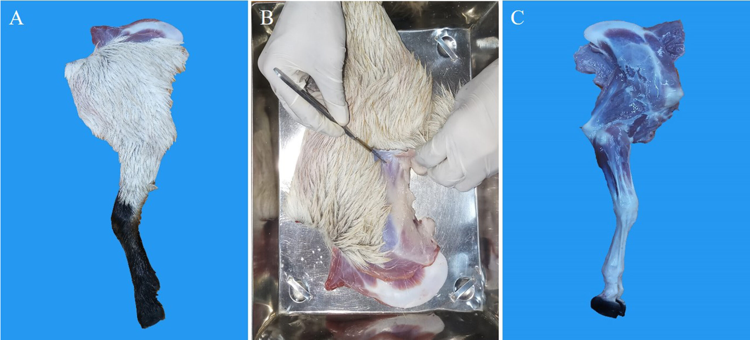 Figure 1. Collection of musculoskeletal specimens of goat: (a) goat forelimb (before removal of skin and fascia); (b) removing skin and fascia; (c) goat forelimb (after removal of skin and fascia) |
Experimental animal and sampling
An indigenous breed of goat was used in this experiment. A clinically healthy experimental animal was purchased from the local market. Euthanasia was performed in a way that minimized fear and anxiety in the animal, and to ensure that the animal cadaver was ethically sourced. The forelimb was then removed from the euthanized goat for further processing (Fig. 1A).
Specimen preparation
At first, the skin was removed carefully to expose the muscle (Figs. 1B & 1C). Then careful incisions were made between the muscle bundles to split them out partially or completely. This was done to ensure proper penetration of the chemicals used in the next steps i.e. formalin, acetone, and glycerin for fixation, dehydration, and immersion, respectively.
Fixation
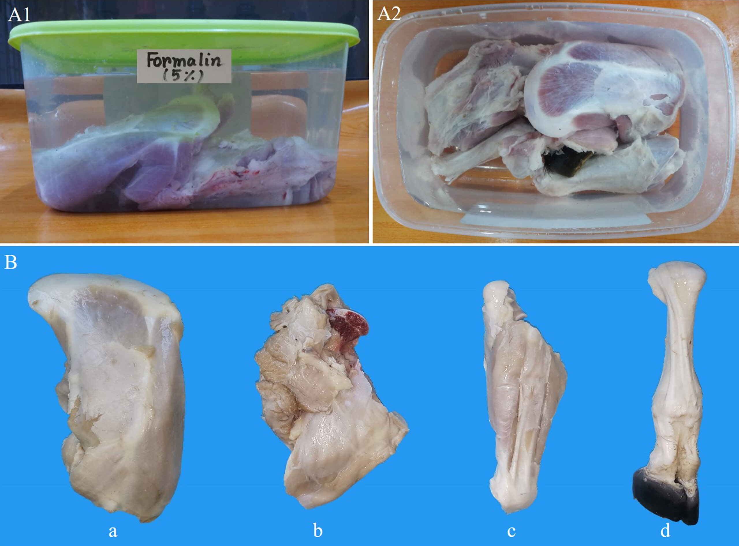 Figure 2. Fixation of musculoskeletal specimens of goat forelimb: (A1, A2) formalin-fixed musculoskeletal parts of goat forelimb; (B) shoulder (a), arm (b), forearm (c), manus (d). The specimens appeared firm and slightly pale in color without any shrinkage |
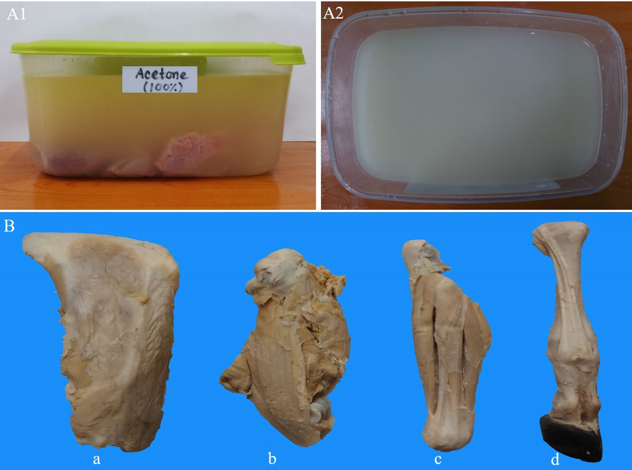 Figure 3. Dehydration of musculoskeletal specimens of goat forelimb with pure acetone. (A1, A2) dehydrated musculoskeletal specimens: shoulder (a), arm (b), forearm (c), manus (d). Dehydrated specimens were firm, with minimal shrinkage (especially shoulder and arm) and good colors |
Formalin is a frequently used fixative in embalming fluid to avoid putrefaction by inhibiting decomposition and deactivating saprophytic microorganisms. In this study, collected specimens were immersed in a 5% formalin bath, and left in the laboratory at room temperature for four weeks for the tissues to become fully saturated and fixed. Such a lower concentration of formalin was used with a view to ensuring the proper fixation of deep muscles (Fig. 2).
Dehydration with acetone
After the completion of the fixation procedure, the specimens were washed with running tap water for 30 minutes to remove the formalin. The water from the surface of the specimens was removed with tissue paper. Afterward, the specimens were kept submerged in pure (100%) acetone for a period of seven days. After seven days, determination of the purity of the acetone solution was performed with a hydrometer to get an idea about the level of dehydration. The specimens were then submerged in another pure (100%) acetone bath for another seven days. After each change of acetone bath, the concentration of acetone was measured. The concentration of acetone was found at 89%, 96%, and > 99% after the first, second, and third weeks of dehydration, respectively. As the concentration of the acetone was found > 99% after the third week of dehydration, it was assumed that the specimens are properly dehydrated. Thus, three changes of pure acetone were needed to complete the dehydration process of musculoskeletal specimens (Fig. 3).
Immersion in glycerin
The acetone was then soaked from the surface of the dehydrated musculoskeletal specimens with tissue paper by applying gentle pressure. Then the specimens were left undisturbed to drain out the acetone for half an hour. Finally, the specimens were fully immersed in a glycerin bath for two weeks for complete impregnation (Fig. 4).
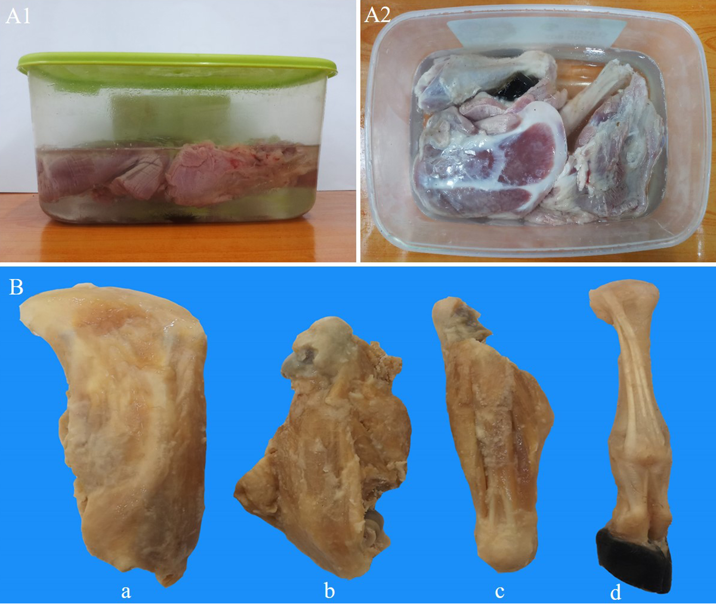 Figure 4. Immersion of musculoskeletal specimens of goat forelimb with glycerin. (A1, A2) glycerin-immersed musculoskeletal specimens, (B): shoulder (a), arm (b), forearm (c), manus (d). Glycerin-immersed specimens were soft, flexible with no further shrinkage. |
 Figure 5. Representative image of desiccation of the musculoskeletal specimens with cornstarch in cloth bag. |
Desiccation with cornstarch
Once the impregnation process was completed, the specimens were removed from the glycerin and left undisturbed for half an hour to drain out. Then the excess glycerin from the surface was removed using tissue paper. Finally, the glycerinated specimens were kept inside a cloth bag containing cornstarch powder (Fig. 5). The cloth bag was then tightly ligated with cotton. Cornstarch powder was dabbed over the cloth bag from the outside.
The specimens were then left in contact with the cornstarch powder for seven days. The specimens were frequently turned within the cloth bag. After seven days, the clumped cornstarch was removed from the surface of the specimen and the previous cornstarch power was replaced with fresh powder. The cornstarch powder was replaced six times during the curing period. Finally, after about two weeks, the starch no longer clumped. The specimens were then carefully taken out of the bag. The specimens were then taken out of the cornstarch powder and the surface was cleaned using a soft brush. The finished specimens were stored at room temperature in a sealed plastic container containing white silica gel granules.
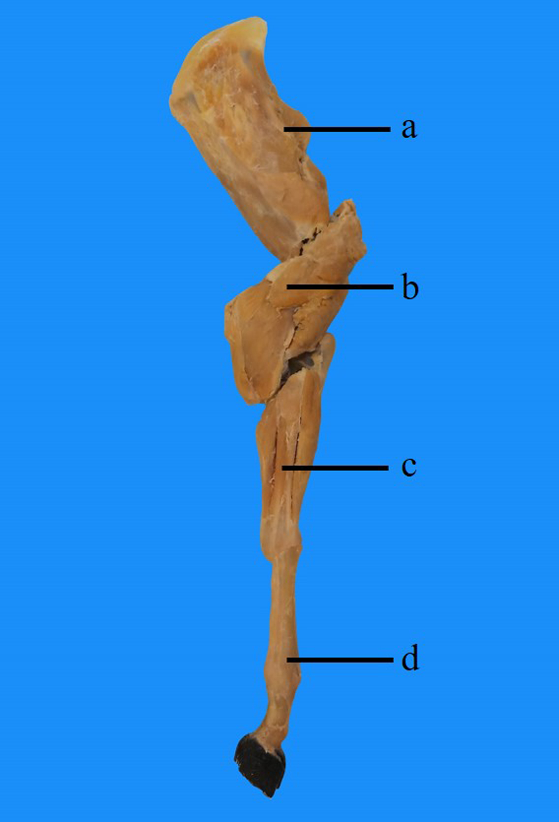 Figure 6. Preserved musculoskeletal specimens of goat: shoulder (a), arm (b), forearm (c), manus (d). The preserved specimens appeared realistic, dry, soft, flexible, and devoid of any significant shrinkage. |
The musculoskeletal specimens (i.e., forelimb) of goat were preserved by an improved method of tissue preservation. The first step, fixation with 5% formalin, gave the specimens a firm and slightly pale appearance without any shrinkage. After dehydration, the dehydrated specimens were firm and had a very negligible amount of shrinkage (especially the shoulder and arm) but produced excellent colors. Following immersion in glycerin, the specimens became soft, and shrinkage appeared after dehydration was gone. The resulting specimens are shown in Figure 6.
After the preservation procedure, the specimens retained their original shape and revealed detailed anatomical structures. Unlike the traditional methods of plastination or tissue preservation, they were more realistic, durable, and soft to touch with higher flexibility. The preserved musculoskeletal specimens appeared clean and there was no objectionable odor. They did not lose any aspects of their physical appearance, such as color, following the plastination process. The specimens had no shrinkage.
The surfaces of the specimens were dry, and they were able to be handled using bare hands. The prepared specimens could be easily stored for a longer period without using any further preservatives. Notably, the approximate cost for preserving musculoskeletal specimens in this method was 51$ (per kg specimen) which indicates the cost-effectiveness of the current method.
Plastination appears to have a bright future in various fields like training and research, as well as public education and culture, all around the globe. It has been gaining attention for its advantages as an educational and research tool (Chandini, 2014). Quick accessibility, easy transportation, and the lack of any need for preservatives have made it an effective educational tool (Rahul et al., 2020). Because of these properties, academics and researchers have been attempting to expand its extension for different educational and research purposes (Steinke et al., 2008). Many anatomy departments now have access to plastinated specimens due to the availability of new quick and risk-free procedures. Plastination provides a unique window into the realm of anatomy for learners, due to lower expenses and the specimens' vibrant look (Pashaei, 2010).
Freshly collected or chemically preserved specimens are commonly used in different anatomy practical classes. Using fresh specimens each time for demonstration is very costly and also questionable from an ethical perspective. Formalin is one of the most frequently used preservatives (Wieding et al., 2015). It is, however, highly toxic to all living beings, regardless of the method of intake, and causes irritation of the skin, eyes, nose, and throat, along with increased tearing (Binawara et al., 2010; Kamruzzaman, 2016). Although formalin was used in our procedure for tissue fixation, it did not require continuous or long-term exposure.
In the dehydration process, alcohol and acetone are commonly used (Steinke et al., 2008; Srisuwatanasagul et al., 2010; Hayat et al., 2018). Dehydration in acetone preserves the natural color of the specimen with little shrinkage, while dehydration in alcohol leads to a darker color and shrinkage (Srisuwatanasagul et al., 2010). In the current technique, the acetone-based dehydration successfully produced good quality specimens. Immersion in glycerin is the next major step, following dehydration. Glycerin had been previously used in different studies to prepare semitransparent to transparent specimens (An et al., 2012; Li et al., 2012). It also can be used for the preservation of different organs or body parts (Silva et al., 2011; Carvalho et al., 2013; Cury et al., 2013). The non-toxic nature, plasticizing and hygroscopic properties, and the very good penetrating power of glycerin into deep structures facilitates tissue preservation (Elnady, 2016). The final step of our tissue preservation technique was desiccating the musculoskeletal specimens with cornstarch. The cornstarch makes the surface of the tissue dry and non-sticky by absorbing the glycerin.
The current method of tissue preservation is much more economic than traditional plastination techniques. To prepare plastinated specimens, it requires approximately 40$ per kg (von Hagens et al., 1987) to 53$ (Dawson et al., 1990) for the chemicals alone. In today’s value, that would be approximately 85$ to 113$, considering the inflation rate. However, the total cost per kg for producing plastinated specimens is much more if the equipment costs are included. In the current method, it cost approximately 51$ per kg, which indicates a 35-55% cost reduction just in terms of the chemicals used. However, this method does not require any specialized equipment or laboratory facilities, which will make it even cheaper and accessible to all.
In conclusion, the authors performed an innovative and comparatively new method of preservation of musculoskeletal specimens of goat developed by Dr. Elnady. It is noteworthy that this work was performed for the first time in Bangladesh. The method was easier in comparison to other methods of plastination such as silicone or sheet plastination, because it did not require any specialized laboratory facilities (i.e., freezer for dehydration, and vacuum impregnation) or expensive chemicals (i.e., polymer, epoxy, or resin). The entire procedure can be done at room temperature.
The prepared musculoskeletal specimens were dry, soft, flexible, and devoid of shrinkage. They were also very realistic, durable, and free from any offensive odor. The prepared specimens produced very good color which was almost the same as the original color of the specimen. The specimens could be stored in an airtight container for long periods without deterioration, and can be handled at any time with bare hands. Due to the advantages of the preservation technique described above, it will be a very effective learning tool for medical and veterinary students.
Acknowledgements:
The authors acknowledge the expert technical assistance of the Departments of Anatomy and Histology,
Bangladesh Agricultural University, Bangladesh. Results from such research will yield a much more economical and healthful tool for preservation of specimens, which can potentially increase overall anatomical knowledge about the musculoskeletal system. Dr. Nasrin Sultana is thankful to the Ministry of Science and Technology, Bangladesh for providing the research fund. (Grant No.: BS-73/MoST-2020-21).
An X, Yue B, Lee JH, Lin C, Han SH. 2012: Arterial anatomy of the gracilis muscle as determined by latex injection and glycerin transparency. Clin Anat 25: 231-234.
https://doi.org/10.1002/ca.21217
Binawara BK, Rajnee CS, Mathur KC, Sharma H, Goyal K. 2010: Acute effect of formalin on pulmonary function tests in medical students. Pak J Physiol 6: 8-10.
Brenner E. 2014: Human body preservation-old and new techniques. J Anat 224: 316-344.
https://doi.org/10.1111/joa.12160
Carvalho YK, Zavarize KC, Medeiros LDS, Bombonato PP. 2013: Evaluation of the glycerin from biodiesel production in the preservation of anatomical parts. Pesq Vet Bras 33: 115-118.
https://doi.org/10.1590/S0100-736X2013000100021
Chandini R. 2014: Plastination. J Pharm Sci Res 6: 271-273.
Cury FS, Censoni JB, Ambrósio CE. 2013: Anatomical techniques in the animal anatomy practice teaching. Pesq Vet Bras 33: 688-696.
https://doi.org/10.1590/S0100-736X2013000500022
Dawson TP, James RS, Williams GT. 1990: How do we teach pathology? Silicone plastinated pathology specimens and their teaching potential. J Pathol 162: 265-272.
https://doi.org/10.1002/path.1711620314
Elnady F. 2016: The Elnady technique: an innovative, new method for tissue preservation. ALTEX 33: 237-242.
https://doi.org/10.14573/altex.1511091
Haizuka Y, Nagase M, Takashino S, Kobayashi Y, Fujikura Y, Matsumura G. 2018: A new substitute for formalin: application to embalming cadavers. Clin Anat 31: 90-98.
https://doi.org/10.1002/ca.23011
Hayat K, Qureshi AS, Rehan S, Rehman T. 2018: Plastination ‐ an innovative preservative technique in anatomy. Trends Anat Physiol 1: 003.
https://doi.org/10.24966/TAP-7752/100003
Islam R, Ayman U, Sultana N. 2021: Histological architectures and biometric characteristics of indigenously plastinated organs of goat. Int J Morphol 39: 759-765.
https://doi.org/10.4067/S0717-95022021000300759
Jordan G, Thomasius R, Schröder H, Wulf JS, Schlüter O, Sumpf B, Maiwald M, Schmidt H, Kronfeldt HD, Scheuer R, Schwägele F, Lang KD. 2009: Non-invasive mobile monitoring of meat quality. J Verbr Lebensm 4: 7-14. DOI
https://doi.org/10.1007/s00003-009-0389-1
Kamruzzaman M. 2016: Formalin crime in Bangladesh: a case study. Eur J Clin Biomed Sci 2: 39-44.
Kim KH, Jahan SA, Lee JT. 2011: Exposure to formaldehyde and its potential human health hazards. J Environ Sci Health 29: 277-299.
https://doi.org/10.1080/10590501.2011.629972
Li J, Shi S, Zhang X, Ni S, Wang Y, Curcio CA, Chen W. 2012: Comparison of different methods of glycerol preservation for deep anterior lamellar keratoplasty eligible corneas. Invest Ophthalmol Vis Sci 53: 5675-5685.
https://doi.org/10.1167/iovs.12-9936
Pashaei S. 2010: A brief review on the history, methods and applications of plastination. Int J Morphol 28: 1075-1079.
https://doi.org/10.4067/S0717-95022010000400014
Piombino-Mascali D, Aufderheide AC, Johnson-Williams M, Zink AR. 2009: The Salafia method rediscovered. Virchows Arch 454: 355-357.
https://doi.org/10.1007/s00428-009-0738-6
Rahul TG, Francis DV, Pandit S, Suganthy J. 2020: Deplastination: preservation of histological structures and its anticipated role in the field of histopathology. Clin Anat 33: 108-112.
https://doi.org/10.1002/ca.23477
Ravi SB, Bhat VM. 2011: Plastination: a novel, innovative teaching adjunct in oral pathology. J Oral Maxillofac Pathol 15: 133-137.
https://doi.org/10.4103/0973-029X.84475
Schoenfeld-Tacher RM, Horn TJ, Scheviak TA, Royal KD, Hudson LC. 2017: Evaluation of 3D additively manufactured canine brain models for teaching veterinary neuroanatomy. J Vet Med Edu 44: 612-619.
https://doi.org/10.3138/jvme.0416-080R
Silva NA, Galvão APO, Fraga KB, de Oliveira RG, Barbosa RF, Campina, RCF, Santos TR, Magalhães CP. 2011: Comparative study between two techniques using a glycerin in the conservation of central nervous system. J Morphol Sci 28: 280-282.
Srisuwatanasagul K, Srisuwatanasagul S, Adirekthaworn A, Darawiroj D. 2010: Comparative study between using acetone and absolute alcohol for dehydration in plastination procedure. Thai J Vet Med 40: 437-440.
https://doi.org/10.56808/2985-1130.2263
Steinke H, Rabi S, Saito T, Sawutti A, Miyaki T, Itoh M, Spanel-Borowskia K. 2008: Light-weight plastination. Ann Anat 190: 428-31.
https://doi.org/10.1016/j.aanat.2008.02.005
Sultana N, Khan MZI, Amin T, Jahan MR, Uddin I. 2019: Preservation of internal organs of goat by an alternative method to plastination. J Plastination 31(1): 14-18.
https://doi.org/10.56507/CMZK7885
Valliyate M, Robinson NG, Goodman JR. 2012: Current concepts in simulation and other alternatives for veterinary education: a review. Vet Med 57: 325-337.
https://doi.org/10.17221/6261-VETMED
von Hagens G, Tiedmann K, Kriz W. 1987: The current potential of plastination. Anat Embryol (Berlin) 175: 411-421.
https://doi.org/10.1007/BF00309677
von Hagens G. 1979: Impregnation of soft biological specimens with thermosetting resins and elastomers. Anat Rec 194: 247-255.
https://doi.org/10.1002/ar.1091940206
Wieding J, Mick E, Wree A, Bader R. 2015: Influence of three different preservative techniques on the mechanical properties of the ovine cortical bone. Acta Bioeng Biomech 17: 137-146.
Yue W, Jin X-B, Pan X-C. 2004: Relationship between indoor air formaldehyde exposure and allergic asthma in adults. Chin J Public Health 20: 904-906. http://www.zgggws.com/en/article/id/16356