Department of Anatomy, Faculty of Health Sciences, University of Durban-Westville, Durban, South Africa
Plastination as a preservation technique, demonstration aid and research tool is well established. The aim of this study was to develop a technique of making solid viscera after latex injection and plastination, transparent. Thirty-five pairs of morphologically normal post-mortem human adult kidneys were harvested en-bloc. These specimens were subjected to various techniques after latex injection and included two types of plastination (P40 and E12), varying combinations of KOH and proteolytic enzyme immersion. Superficial transparency was achieved for only 1-2 mm in the en-bloc samples. Acceptable transparency was achieved only in coronally sectioned samples. The technique of latex injection, immersion in 10% KOH (6 days), slicing, dehydrating and subjecting to the E12 plastination technique produced the best results thus far with acceptable transparency of the solid visceral tissue. In the total series of 35 pairs of kidneys the quest for transparency still remains elusive.
Transparency, Sections, Immersion
Professor KS Satyapal, Head, Department of Anatomy, Faculty of Health Sciences, University of Durban-Westville, Private Bag X54001, Durban 4000, South Africa. Telephone: 27 31 204 4195 / Fax: 27 31 204 4890. Email: kapil@pixie.udw.ac.za

![]()


Plastination as a preservation technique, demonstration aid and research tool is well established. While many investigators continue to improve preservation techniques, one of the overwhelming challenges that the technique of plastination faces is an ability to select certain anatomical structures to be highlighted and facilitate transparency of the surrounding tissue. Being able to replicate through plastination, for example, a renal angiogram with the parenchyma intact but transparent will contribute enormously to teaching and research since once more we will be dealing with "the real thing." Thus far the E12, P35 and P40 techniques of plastination were used to produce transparent or semi-transparent slices of tissue but were not utilized for whole organs (von Hagens, 1985). The P40 as well as the P35 plastination techniques were used for the production of thin (4,6 or 8 mm), opaque slices of brain tissue (Barnett, 1997). However, the literature reviewed does not describe success in the quest for selective whole organ transparency but does record plastination of a cleared fetus to show vascularization of ossifying bone (Haffajee, 1996). The aim of this study was to develop a technique of making solid viscera after latex injection, transparent.
Thirty-five pairs of morphologically normal post- mortem human adult kidneys were harvested en-bloc. The arterial, venous and uretero-pelvi-caliceal systems were perfused with de-aerated warm water, gently massaged and on obtaining a clear perfusate, immersed in 5% formalin (2000ml) for 24 hours at 18°C. The three systems were injected with different colored rubber latex: red-arterial (100ml), blue-venous (150 ml) and yellow-pelvi-caliceal (50ml), according to the method described by Tompsett in 1970 (figure 1). The rubber latex was supplied by: Genkem Pty Ltd, PO Box 120121, Jacobs, Durban, 4000, South Africa. Specimens were left to cure in absolute alcohol for 3 days at 18°C. Thereafter, the kidneys were subjected to a series of different random procedures and grouped as follows (Table 1).
These were:
The results according to the groups subjected to the different techniques (table 1) were :
| Group and Sample size
(en-bloc kidney pairs) |
Technique used | Results |
| a) 10 En-bloc renal specimens
|
Rubber latex injection + E12 plastination | Minimal superficial transparency achieved for only 1-2mm |
| b) 7 Coronal slices | Rubber latex injection + E12 plastination | Acceptable but patchy transparency |
| c) 4 En-bloc renal specimens | KOH immersion | Poor transparency : less than 2mm attained with tissue maceration |
| d) 2 En-bloc renal specimens | Proteolytic enzyme (Pepsin) immersion | Poor transparency : less than 2mm attained with tissue maceration |
| e) 2 En-bloc renal specimens | KOH + Pepsin immersion | Poor transparency : less than 2mm attained with tissue maceration |
| f) 8 Coronal slices | P40 and E12 plastination | Poor transparency (less than lmm) with P40 technique; better quality transparency (less than 3mm) with E12 technique |
| g) 2 Coronal slices | KOH immersion + E12 plastination | Acceptable transparency (preferred technique which has potential for best results) |
Group a
En-bloc renal specimens (rubber latex injection + E12 plastination):
Minimal superficial transparency, only between 1-2 mm of the renal parenchyma was attained. However, the vessels (rVC, aorta and renal artery and vein) and the pelvis and ureter were observed to be transparent.
Group b
Coronal slices (rubber latex injection + E12 plastination):
Acceptable but patchy transparency of the renal parenchyma was achieved. It was noted that the periphery remained relatively opaque. Partial visualisation of the intra- renal vessels was observed.
Groups c,d,e
En-bloc renal specimens (KOH; Pepsin; KOH + Pepsin immersion):
Poor transparency of less than 2 mm was attained together with tissue maceration.
Group f
Coronal slices (P40 and E12 plastination):
Poor transparency (less than 1 mm) was obtained using the P40 technique. The E12 technique was minimally superior, achieving less than 3 mm transparency.
Group g
Coronal slices (KOH immersion + E12 plastination):
Acceptable transparency was obtained with this technique which yielded the best results in this experimental series. However, the transparency of the parenchyma was also partial in areas. Differentiation between the medulla and cortex was observed. In addition, a finer visualisation of the intra-renal vasculature was noted.
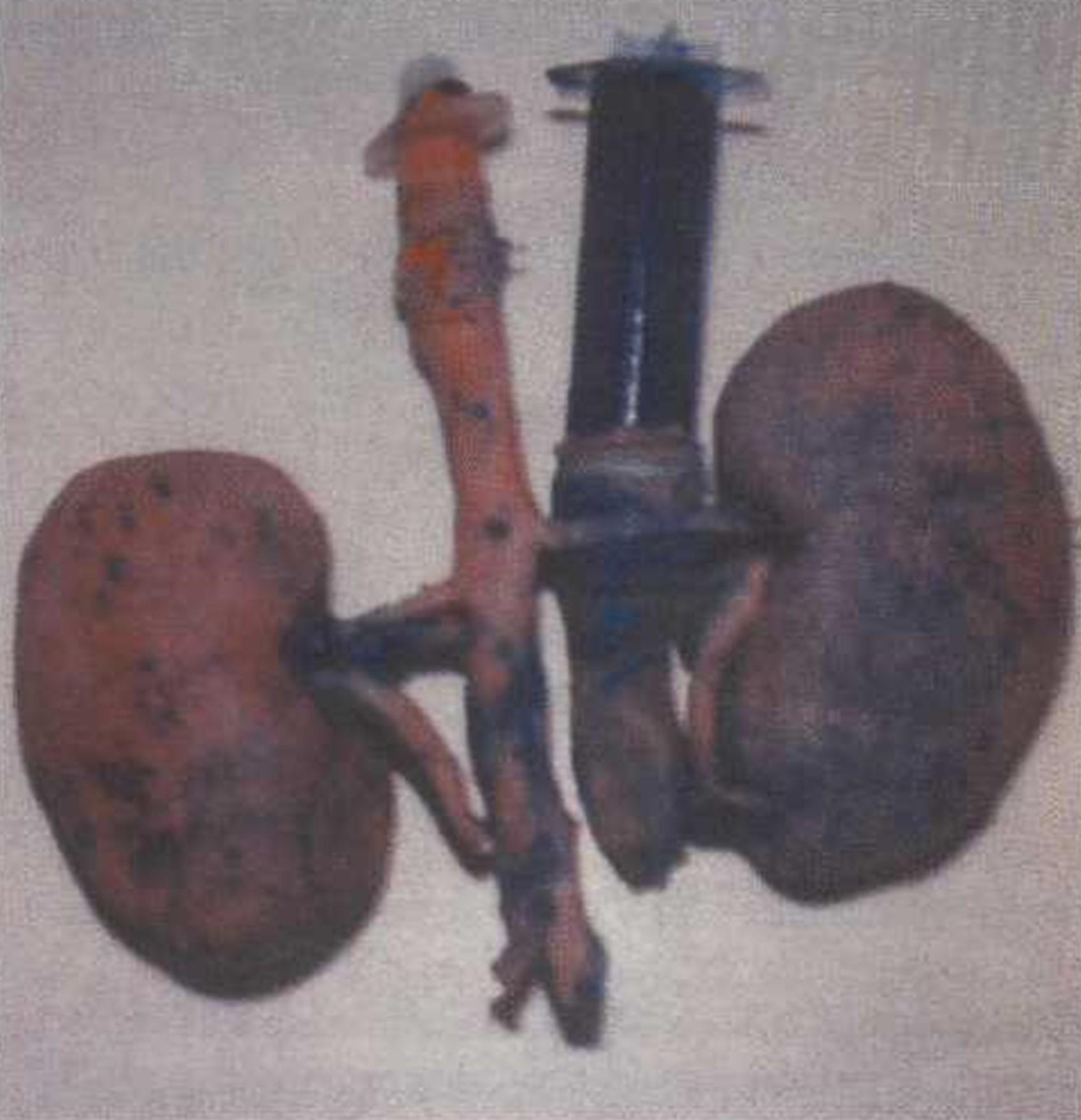 Figure 1. Uretero-pelvi-caliceal system injected with different colored latex in en-bloc specimen. |
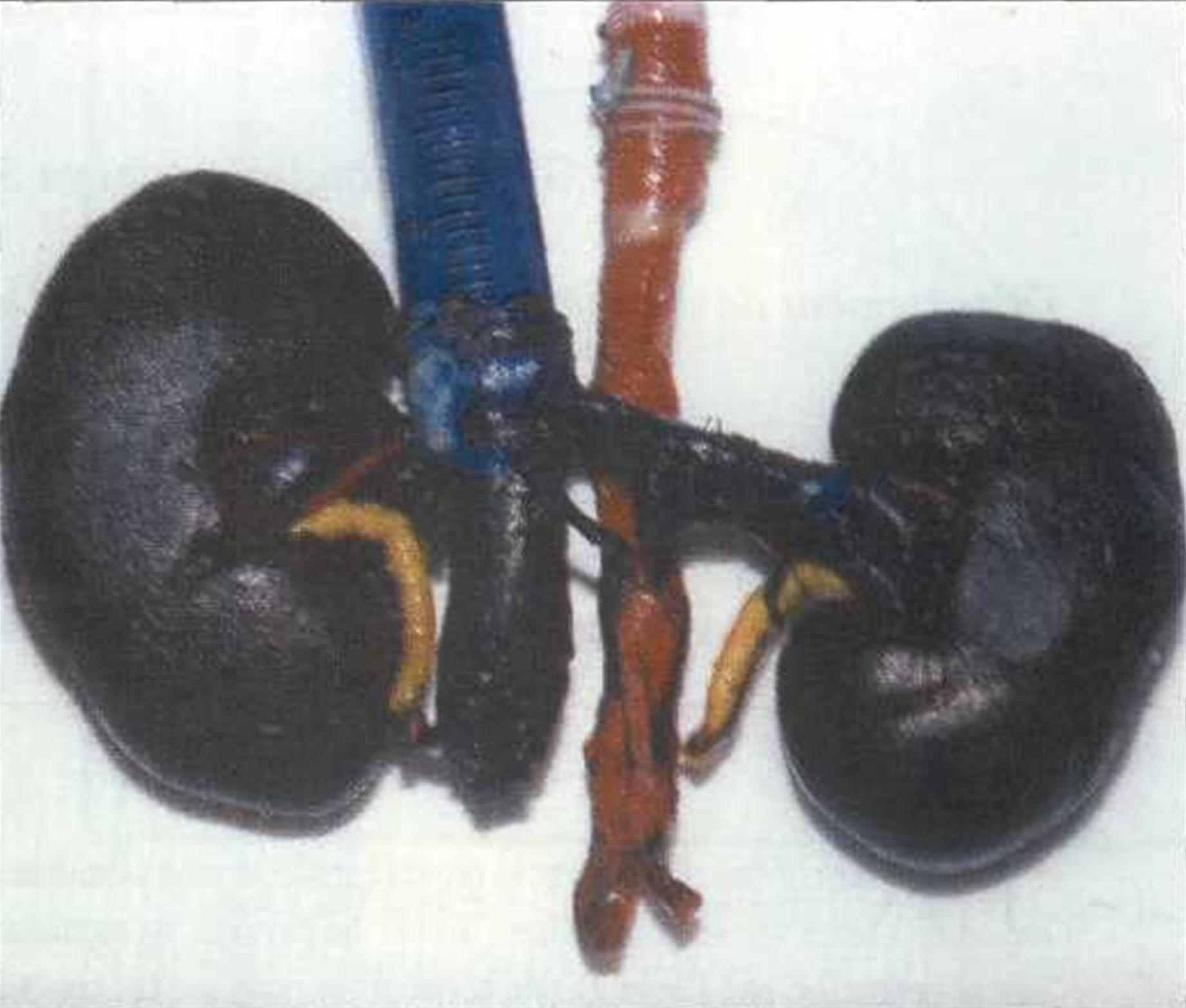 Figure 2. Minimal superficial transparency : between 1-2 |
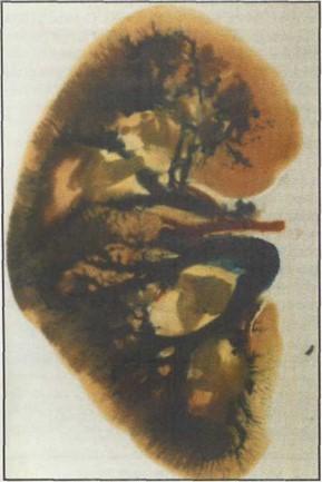 Figure 3. Coronal slices: acceptable but patchy transparency was achieved (Group b). |
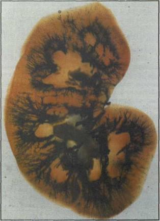 Figure 4. Coronal slices : acceptable transparency |
The results of our experiment clearly indicate that significant further research is required to attain the desired level of transparency in a whole organ after injection of vessels with rubber latex. It is clear that Groups a-f hold little promise in attaining these results since the degree of transparency was minimal as evidenced in figures 2 and 3. In addition, Groups c, d and e were clearly unacceptable since tissue maceration also occurred. Our results confirm that tissue slices lend themselves better to transparency using plastination.
The technique of latex injection, immersion in 10% KOH (6 days), slicing, dehydrating and plastination according to the E12 technique has produced the best results thus far with acceptable transparency of the solid visceral tissue. In the total series of 35 pairs of kidneys the quest for transparency still remains elusive. The researchers continue to experiment with combinations of various techniques in the quest for transparency in plastination.
Barnett RJ: Plastination of coronal and horizontal brain slices using the P40 technique. J Int Soc Plastination 12 (1): 33-36, 1997.
https://doi.org/10.56507/YJVS5787
Haffajee MR : Plastination of a cleared fetus to show vascularization of ossifying bone. J Int Soc Plastination 10 (1): 26-27, 1996.
https://doi.org/10.56507/FZUI4078
Tompsett DH : Anatomical techniques. 2nd ed. Livingstone, Edinburgh, 1970.
von Hagens G: Heidelberg Plastination Folder. Anatomisches Institut, Universitat Heidelberg, Heidelberg, Germany, 1985.