St George’s, University of London, London, UK
The Journal of Plastination, as we know it today, came into being as the “Journal of the International Society for Plastination” thirty-one years ago, in January 1987, under the distinguished Editorship of Dr Harmon Bickley . The cover, a simple white with red lettering, featured an axial image of the human abdomen. It is not clear where this image came from, as there were no figures at all inside this first issue. However, one of the papers within it details the procedure for plastination of whole-body slices with Biodur® S10 or epoxy: “Sectional anatomy is a valuable approach to the acquisition of an understanding of body structure. Before our use of plastinated slices, it had been neglected for many years”
journal
Philip J. Adds, MSc, SFHEA, FAS, FIBMS
Biomedical Sciences (Anatomy)
St George's University of London
Cranmer Terrace
London SW17 0RE
Phone: +0208 725-5208
E-mail: philadds.anatomy@gmail.com

![]()


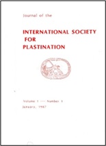 Figure 1. Front cover of Volume 1, Number 1, January 1987 |
The Journal of Plastination, as we know it today, came into being as the “Journal of the International Society for Plastination” thirty-one years ago, in January 1987, under the distinguished Editorship of Dr Harmon Bickley (Fig 1). The cover, a simple white with red lettering, featured an axial image of the human abdomen. It is not clear where this image came from, as there were no figures at all inside this first issue. However, one of the papers within it details the procedure for plastination of whole-body slices with Biodur® S10 or epoxy: “Sectional anatomy is a valuable approach to the acquisition of an understanding of body structure. Before our use of plastinated slices, it had been neglected for many years” (Lischka & Prihoda, 1987).
The presence of this image on the cover is highly significant, and demonstrates the importance (and relevance) of sheet plastination to sectional anatomy and, hence, to healthcare technology. While CT and MR were still relatively new in the eighties, they have since come to dominate medical imaging, and, crucially, depend on the ability of medical practitioners to interpret sectional images. Sectional anatomy is now considered to be an integral part of medical education: even back in the eighties, the Journal was ahead of the field.
Five papers were published in issue 1, mostly describing technical aspects of tissue preservation and plastination; one paper, however, recognised the potential of plastination in research: “Complete Examination of Mastectomy Specimens Using Sheet Plastination with Epoxy Resin”, which had among its authors the inventor of plastination, Gunther von Hagens (Guhr et al., 1987).
The very first paper in the new journal was by Karine Oostrom from Utrecht in The Netherlands: “Fixation of Tissue for Plastination: general principles”, which discussed different methods of fixation, color preservation, colour injection, health hazards, and employee safety (Ostrom, 1987). Ostrom described the personal protective equipment (PPE) worn in the plastination lab in Heidelberg: “rubber gloves, plastic aprons, and goggles or gas masks.” Ostrom continued: “Those of you who attended the Third International Conference on Plastination in San Antonio will certainly recall the slide in which three young ladies modelled these fashionable accessories, and nothing else…The editor was adamant that we omit this illustration, however it would have served to show that even fixation can be fun.” It is good to note that high editorial standards were already in place!
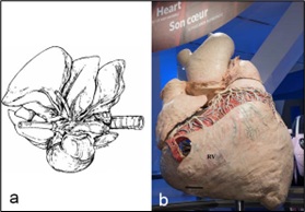 Figure 2. a) Canine heart-lung specimen (Henry, 1987); b) blue whale heart (Miller et al., 2017) |
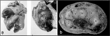 Figure 3. a) Heart valve specimen (Baptista & Conran, 1989); b) wrist section (Baptista et al., 1989) |
The second issue followed in the same year, again with five papers focussing mainly on technical issues. Notable authors in issue 2 include Dr Robert Henry, who contributed a paper on the plastination of hearts and heart-lung specimens, using canine specimens (Henry, 1987). Thirty years later, Dr Henry was among the authors of a paper describing heart plastination on a much larger scale: the salvage and preservation of a blue-whale heart: “The Challenges of Plastinating a Blue Whale (Balaenoptera musculus) Heart “(Miller et al., 2017). It is interesting to compare the image that accompanied the 1987 paper, (which was in fact the first anatomical image ever to appear in the Journal), with the image of the final specimen that accompanied the 2017 paper (Fig. 2). These images help to show how far plastination (and the Journal) have come in the last 30 years. Progress indeed.
The author affiliations from Volume 1 show that all contributing authors were from the early centres of plastination, one each from Utrecht, Vienna, and Heidelberg, and the rest from the USA. This narrow geographical range reflects the limited reach of the emergent technology of plastination at that time.
The next significant develop in the development of the journal came 2 years later, in 1989, with the appointment of Bob Henry as Editor; Harmon Bickley became Executive Director of the Society. Volume 3 (1989) was single-issue only, and included, for the first time, abstracts from meetings of the International Society for Plastination: the Fourth International Conference (held in 1988 at Macon, Georgia, USA) and the inaugural Interim Meeting, held a year later in Knoxville, Tennessee. Volume 3 also heralded the arrival in print of another significant figure, Dr Carlos Baptista, with two papers focussing on clinical and applied human anatomy, “Plastination of the heart: preparation for the study of the cardiac valves” (Baptista & Conran, 1989), and “Plastination of the wrist: potential uses in education and clinical medicine” (Baptista et al., 1989) (Fig. 3), the latter again focussing on sectional anatomy. As the authors put it: “These specimens provide an excellent tool for teaching anatomy and pathology, for patient education, and potentially as an augmentation to MRI (magnetic resonance imaging) and CT (computer tomography) analysis” (Baptista et al., 1989).
The next step in the evolution of the Journal came with Volume 4, another single-issue volume, published in the fall of 1990. For the first time, an Editorial board appeared on the first page, consisting of Drs Carlos Baptista, Harmon Bickley, and P. Tom Purinton. Bob Henry continued as Editor, and Harmon Bickley as Executive Director of the Society. Volume 4 contained the usual eclectic mix of research and technical papers. Notable among them was a paper by Lane, continuing the theme of sectional anatomy “Sectional anatomy: standardized methodology” (Lane, 1990), with an x-ray image showing levels of axial sections of the body (Fig. 4) and color images of plastinated body sections (Fig. 5).
It is interesting to compare these early papers on sectional plastinates with the “Visible Human Project”, which claimed to have revolutionised the study of anatomy: “The National Library of Medicine (NLM) introduced the Visible Human Project (VHP) in November 1994 and in doing so revolutionized our ability to view and understand human anatomy.” (https://infocus.nlm.nih.gov/2014/12/31/the-visible-human-project-at-20/), emphasis added). On the contrary, it could be claimed that plastination had already achieved this, nearly a decade earlier!
Another paper in Volume 4 discussed the potential for using plastinates in the construction of holographic images (Myers and Bickely, 1990). Holography was described at the time as “a solution in search of a problem” (ibid.), because it had thus far fulfilled only a small part of its potential. Despite speculation about the potential for holograms in medical education, the technology never really seemed to take off. Virtual reality is now the only game in town. Try typing “plastination hologram” into Google images nowadays, and the only relevant image likely to appear is Gunther von Hagens with a hologram of a plastinated couple during intercourse (Fig. 6).
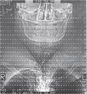 Figure 4. Levels of sectioning for axial body slices (Lane, 1990) |
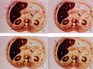 Figure 5. Images of plastinated body sections |
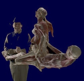 Figure 6. Gunther von Hagens with a hologram of a couple during intercourse |
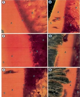 Figure 7. Photomicrographs of sheet plastinated human knees, showing a control and a series of slices showing degenerative changes (Graf et al., 1992) |
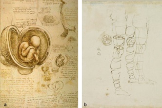 Figure 8. “First known examples of cross-sectional anatomy by Leonardo da Vinci (1452 – 1519) of the pregnant uterus (a) and the lower limb (b)“ (Weiglein, 1993) |
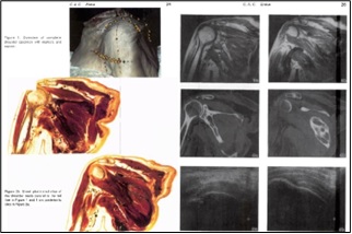 Figure 9. The original shoulder specimen, E12 plastinated slices and corresponding MRI, CT and US images (Entius et al., 1993 |
 Figure 10. The dawn of a new era: The International Society for Plastination is now an official body. |
Volume 6 came out in 1992, another single-issue volume, which contained a paper from Graf et al., “Early Morphological Changes in Chondromalacia Patellae in Humans - Demonstrated With The Plastination Method”, which described how epoxy sheet plastination was used on human patellas to investigate, at the microscopic level, changes in the chondral and subchondral areas. It was reported, for the first time, that “the origin of the idiopathic chondromalacia was detected in the subchondral area and not the cartilage as previously thought.” These changes had not previously been seen with arthroscopy, but demonstrated how plastination can bridge the gap between the macro- and the microscopic, and shows the application of plastination to both research and clinical medicine (Fig. 7) (Graf et al., 1992).
The Journal developed further in 1993, with changes to the editorial team that were announced in Volume 7. Dr Robert Henry remained as Editor, but R. Dale Ulmer, from the College of Medicine, Mobile, Alabama, was listed as Editor-elect. The Editorial Board remained as before, but, as evidence of the growing worldwide reach of plastination, special Journal Correspondents were listed: for Canada, R. Blake Gubbins (Queen’s University, Ontario); for Europe, Margit Rokel (St Leon-Rot, Germany); and for the Far East, Robert Boyes (Queensland, Australia).
Volume 7 also contained a paper from another author who was to go on to become a prominent and distinguished member of the ISP, Andreas Weiglein: “Plastinated Brain Specimens in the Anatomical Curriculum at Graz University”. In this paper, Dr Weiglein described the use of P35 plastinated brain slices in neuroanatomy teaching, but he also draws comparisons between modern-day sectional anatomy and the anatomical drawings of Leonardo da Vinci (1452 – 1519) “the first known examples of cross-sectional anatomy” (Weiglein, 1993) (Figure 8). This was the first paper in the Journal to discuss the history of anatomy in relation to modern anatomical techniques.
In the same issue, a paper by Entius et al. brings state of the art sectional anatomy, medical imaging, and plastination together. In “A New Positioning Technique for Comparing Sectional Anatomy of the Shoulder with Sectional Diagnostic Modalities: Magnetic Resonance Imaging (MRI), Computed Tomography (CT) and Ultrasound (US)” the authors describe how skin markers can be used on anatomical specimens to define planes of section. After MRI, CT and ultrasound images were obtained, the specimen was frozen and sectioned at 2 mm thickness. The slices were then plastinated using the E12 technique, giving sections that exactly matched the MRI, CT and ultrasound images (Entius et al., 1993) (Fig. 9).
Volume 8, in 1994, came with a change in the Editorial team. Dale Ulmer took over as Editor, and Bob Henry joined the new-look Editorial Board, along with Vincent DiFabio, Bill Richeimer, and William A. Gardner Jnr. “Preparation Support”, another innovation, was provided by Betty Clark and Rosemary Farmer.
Volume 9 (1995), with a suitably sunny cover picture (Fig. 10), announced the dawn of a new day for plastination, with the founding of the International Society for Plastination as an official body. At the historic 4th Biennial Meeting, in Graz, Germany, Bylaws and a Constitution for the ISP were written and adopted, and a slate of Officers were proposed and elected, and the Journal carried, for the first time, Letters from the newly-elected President, Bob Henry, and the Editor, Dale Ulmer.
The first-ever Editor’s letter, from Dale Ulmer, carried this message: “I challenge each plastinator to contribute one article yearly to our journal and help us advance our organizational goals. As we learn – we grow.” A message just as relevant today as it was in 1995.
The 8th International Conference on Plastination (the 5th Biennial Meeting of the International Society for Plastination) ventured, for the first time, to the Southern Hemisphere, to the University of Queensland, Brisbane, in Australia. Confusingly, the cover of Volume 10 featured a photograph showing the Sydney harbour bridge and the Sydney Opera House. The abstracts from the meeting were published in this issue, including “The Use of Silicone Plastinated Specimens for Light and Electron Microscopy” (Grondin et al., 1996), which contained the following cryptic message: “if you can find mistakes in this publication, please consider they are there for a purpose. We publish something for everyone, and some people are always looking for mistakes”!
In Volume 11 (1996), an article by Sharon Korbeck entitled “A Pharaoh’s Farewell: the Making of a Mummy” was reprinted with the permission of The National Funeral Directors Association. In his Editor’s Letter, Dale Ulmer wrote “While this process is not true plastination, it is, however, a forerunner to the now popular process that we as Plastinators now use. From time to time, I believe it is good to examine and see the yester years”. This passage reminded me of something Craig Goodmurphy had said at the 15th Biennial Business Meeting of the International Society for Plastination, (Honolulu, July 2010): “…the objective of the Journal of Plastination [should] be expanded to provide a medium for the publication of scientific papers dealing with all aspects of preservation of biological specimens including plastination, sectional anatomy and other anatomical techniques.” This is a very laudable aim, which had the backing of the meeting and the Editorial Board. In fact, as has been shown, the Journal has been doing that all along, but it is good to be reminded from time to time of the importance of the history, and range, of anatomical techniques.
Volume 12 brought the first decade to a close in fine style. The cover of Issue 1 featured stunning colour photographs of stained plastinated brain slices (Fig. 11) from Suriyaprapadilok and Withyachumnarnkul’s (1997) paper “Plastination of Stained Sections of the Human Brain: Comparison Between Different Staining Methods”. Compare this to the cover of Volume 1! There had also been a change in the Editorial team, with Gilles Grondin taking over as Editor, supported by an expanded, international Editorial Board, of Pamela Arnold, Harmon Bickley, Robert Henry, Steven Holladay, Larry Janick, Tage N. Kvist, William Richeimer and Bill Wise from the USA, Russell Barnett from New Zealand, Régis Olry from Canada, and Andreas Weiglein, from Austria.
Issue 2 of Volume 12 was published in October 1997, and included a typically wide-ranging mix of papers, discussing both cutting-edge research, and the rich heritage of anatomy. “Submacroscopic Interpretation of Human Sectional Anatomy Using Plastinated E12 Sections” (Cook and Al-Ali, 1997), expanded on the possibilities offered by E12 plastination in anatomy education “The E12 process … has in effect filled a void in undergraduate teaching. Students are provided with a clear, unimpeded overview of the planes of the body seen with a whole section…. providing a firm link between macroscopic and microscopic anatomy” (Fig. 12).
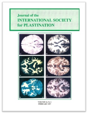 Figure 11. Stained brain slices from Suriyaprapadilok |
 Figure 12. E12 sections. Clockwise from top left: coronal head, right eye (magnified), axial thorax section, thoracic wall (magnified), coronal shoulder section, nasal cavity (magnified). From Cook and Al-Ali (1997). |
 Figure 13. Dissected (L) and injected (R) lymphatic vessels by Rausch, 1665. From Olry and Motomiya (1997) |
While Cook and Al-Ali were looking to the future, Olry and Motomiya (1997) looked back to the Renaissance and beyond, with their paper “Paolo Mascagni, Ernest Alexandra Lauth and Marie Philibert Constant Sappey on the Dissection and Injection of the Lymphatics”. They describe how the lymphatics were “discovered by chance, misunderstood for a very long time, … the subject of much controversy up to the early twentieth century, when their accurate description was deemed necessary to promote advances in oncology” (Fig. 13). It is remarkable that the lymphatic system, though apparently first described (albeit inaccurately) by Erasistratus around 250 BC, was a source of continued controversy up to the 20th century (the role of the thymus, for example, was not fully understood until the 1960’s).
That seems a fitting way to conclude this survey of the first decade of the Journal of Plastination – remembering the past, while looking to the future. In closing, I would like to take the opportunity to repeat the words of Dale Ulmer, in the very first Editor’s letter “I challenge each plastinator to contribute one article yearly to our journal and help us advance our organizational goals. As we learn – we grow”.
Baptista CAC, Conran PB: 1989: Plastination of the heart: preparation for the study of the cardiac valves. J Int Soc Plastination 3: 3-7
https://doi.org/10.56507/PEXL8984
Baptista CAC, Skie M, Yeasting RA, Ebraheim N, Jackson WT. 1989: Plastination of the wrist: potential uses in education and clinical medicine. J Int Soc Plastination 3: 18-21
https://doi.org/10.56507/XENF9035
Cook P, Al-Ali S. 1997: submacroscopic interpretation of human sectional anatomy using plastinated E12 sections. J Int Soc Plastination 12(2): 17-27
https://doi.org/10.56507/XICY2283