1Department of Animal Health (Parasitology and Parasitic Diseases)
2Department of Anatomy and Comparative Pathological Anatomy, Regional Campus of International Excellence
University of Murcia, Murcia, Spain
Plastination is a method of preserving biological tissue in a dry, odorless state, avoiding the use of traditional chemicals, some of them carcinogenic, such as formaldehyde. In recent years, plastination has been introduced in many anatomy departments of human and veterinary medicine, where plastinated specimens are used as a teaching tool for gross anatomy. There are very few references in the literature describing the plastination of parasitic specimens. Formalin-fixed Oestrus ovis larvae, stages L2 and L3, were dehydrated in acetone, and plastinated with the standard Biodur® S10 protocol. Plastinated larvae suffered a collapse of their structures, with a macroscopic deformation of their appearance. In a second test, making an incision in the ventral part of the cuticle allowed us to obtain, for the first time, plastinated larvae of Oestrus ovis, preserving the morphological characteristics that these specimens had before the plastination process.
Cuticle, Oestrus ovis, Parasitology, Plastination, S10 technique
Moisés Gonzálvez, Department of Animal Health (Parasitology and Parasitic Diseases), Regional Campus of International Excellence “Campus Mare Nostrum”, University of Murcia, Spain. Email: moises_vet@hotmail.com

![]()


Nowadays, conservation of parasitic specimens for teaching or research is necessary. However, the most commonly used preservatives (formaldehyde and alcohol) have disadvantages, such as toxicity, carcinogenicity, odor, constant preservative maintenance, storage space, etc.
The technique of plastination (Von Hagens et al., 1987) is generally presented as the most recent and important option for conservation of biological material, which is demonstrated by its rapid expansion in recent years. Plastination consists of processes whereby tissue fluids and fat are slowly replaced by a curable polymer under vacuum conditions. This technique allows us to obtain clean, dry, resistant preparations of unlimited duration, which can be examined without gloves or any other type of protective equipment, and do not require any special treatment or storage conditions. It also avoids the daily exposure of students and teachers to harmful products because they are free of toxic substances, such as formaldehyde, phenol, alcohols, etc. (McLaughlin, 1994; Swenberg et al., 2013).
Plastinated organs enhance the quality of the teaching-learning process at different educational levels in secondary (biology, zoology), and university (anatomy, pathological anatomy, etc.) (Latorre et al., 2007). However, there are very few studies that refer to the conservation of parasites using plastination techniques for teaching or research purposes (Asadi & Mahmodzadeh, 2004; Kocevski et al., 2010; Essa et al., 2014). Preliminary results from studies on plastination techniques applied to parasitology show that each parasite from each of the different taxa needs a specific plastination protocol, in order to preserve their particular morphological and structural characteristics (Grondin et al., 1994)
The objectives of this study were: 1) to evaluate the morphology and morphometry of Oestrus ovis larvae, stages L2 and L3, preserved with the standard S10 plastination protocol; and 2) to establish the necessary changes in the protocol to improve the final results of the plastinated specimens.
Twenty Oestrus ovis larvae from the collection of the Animal Health Department, University of Murcia, Spain, were used in this study, divided in two assays (Fig. 1). For the first trial, ten formalin-fixed larvae (two immature or L2, and eight mature or L3) were used. All larvae were individually evaluated to identify and discard
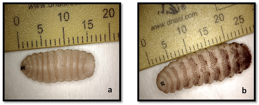
Figure 1a and 1b
specimens with anomalous features, or structural alterations. Each specimen was individually measured to obtain baseline morphological parameters, in order to be able to evaluate changes during each phase of the process.
For this evaluation, a photomicroscopy system was used (Leica EZ Camera 2.4.1) and a specific morphometry software (LAS EZ 2.1.0). The parameters measured were: length (cranio-caudal, maximum body width, minimum width (distance between antennal lobes), and weight. These parameters were recorded after fixation, dehydration, impregnation, and curing.
The standard Biodur® S10 silicone technique was used (DeJong & Henry, 2007). Dehydration was carried out by freeze substitution with acetone (-25° C); forced impregnation was carried out at -25° C under vacuum using silicone S10 and catalyst S3 (100:1) as the reaction mixture. Curing was performed at room temperature using vaporized S6 cross-linker.
Histological cross sections from plastinated larvae were obtained to determine potential structural alterations associated with the process. One mature specimen, and other immature larvae from each assay were used for the histological study. As a control group, different to the two assays, a fixed mature larva and other fixed immature larvae were used. A de-plastination protocol (Grondin et al., 1994) was necessary in order to obtain histological sections from the plastinated larvae.
A second experiment was necessary to improve on the results obtained from the first test. In this second assay, ten additional larvae (five immature L2, and five mature L3) were used. The conventional protocol was modified, making a longitudinal incision of approximately one centimeter on the belly of the parasite, to prevent the cuticle becoming a barrier during acetone and impregnation mixture exchange. As in the previous experiment, the morphometric parameters were monitored to analyze potential structural variations. Parameters evaluated in both experiments were analyzed for statistical differences using a non-parametric Wilcoxon test. Significance was set to p <0.05.
First assay
Plastination protocol: Dehydration by cold substitution of acetone took approximately 5 days, with a total of three baths (Table 1). From the 5th day, the purity of the acetone did not fall below 99.8%. Even so, the specimens were kept for 6 more days in the last bath, to compensate for the possible retardation of dehydration by the cuticle. Impregnation in the S10 + S3 solution did not present any problems, and it took 5 days to reach a final pressure of 5 mmHg, and to achieve the cessation of bubbling of acetone vapor. At the end of impregnation, the samples were removed from the impregnation chamber to room temperature for 24 hours before initiating the polymerization process. The Oestrus ovis specimens were left in the curing chamber while removing excess silicone from the surface. During the curing phase, the organisms were removed from the chamber and stored at -20° C, when excessive surface silicone was not able to be controlled, such as at night. In total, the specimens remained in the polymerization chamber for 24 days (Table 1).
| Assay | Dehydration (days) |
Impregnation (days) | Cured (days) |
| first assay | 11 | 5 | 24 |
| second assay | 7 | 15 | 9 |
Table 1. Differences in the duration of different stages in both assays.
Morphological results: The results obtained in this first test were not satisfactory. Only one specimen
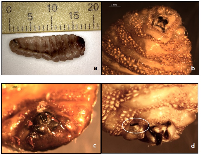
Figures 2a, 2b, 2c and 2d
retained the appearance of its initial state. During dehydration and impregnation, no apparent morphological problems occurred, but during the curing process, the specimens suffered a severe collapse, with alteration of the normal anatomy (Fig. 2a). Two of these plastinated parasites had excessive macroscopic disruption of the
cuticular structures, especially in the rows of spines that border each segment of the larva (Fig. 2b). In addition, one of the specimens showed a deposit of multiple small granules of reddish coloration, approximately half a millimeter in diameter, corresponding to silicone waste (Fig. 2c). After curing, it was possible to observe the existence of damaged and/or broken structures, such as the buccal hooks of these parasites (Fig. 2d). Some defects began to appear in some individuals during the early stages of processing, especially at the end of dehydration, where the fragility of the organisms was evident.
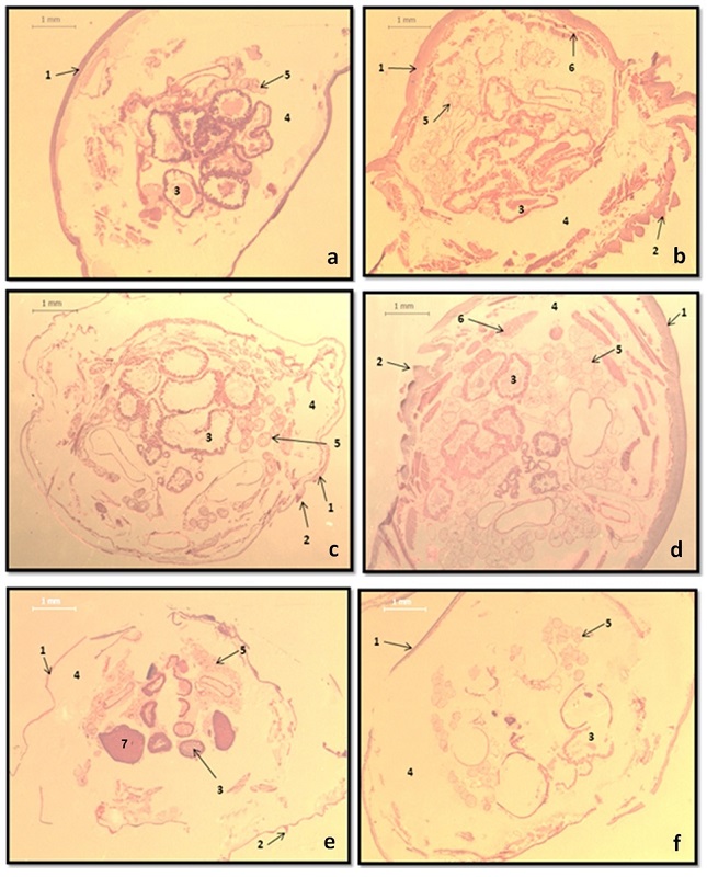
Figure 3a, 3b, 3c, 3d, 3e and 3f
The histological study, comparing non-plastinated with plastinated larvae, was designed to determine the possible causes of the morphological alterations resulting from the plastination process (Fig. 3). Histological evaluation of the fixed larvae permitted appreciation of the great thickness of its cuticle (340 μm). Histological images of larvae (L1 and L2) before and after plastination did not show any other alterations that could explain the reason for the collapse that the structures suffered during processing.
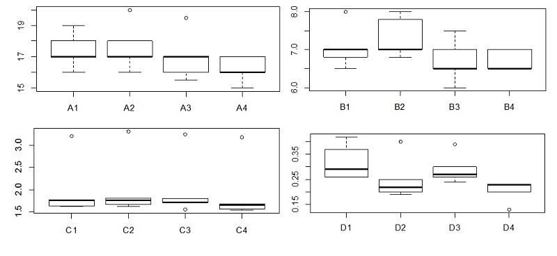
Morphometric results: Table 2 shows the variations of the morphometric parameters of the larvae between each phase, and in the total plastination process, expressed as a percentage of the initial value for all variables studied (length, width and weight). As shown in the table, the final result was a decrease in length of both types of larvae, together with an increase in the maximum width of the individuals. However, there was an increase in the minimum width of the L2, unlike the L3, which presented a decrease in this parameter. These variations in width corresponded to morphological deformations on the surface of these parasites, as a consequence of their collapse after curing. The variable that showed the greatest decrease, with significant differences, was the weight, which could be attributed to a failure in dehydration or impregnation. Graph 1 (L2) and Graph 2 (L3) show the variation of the four variables measured during processing of the samples.
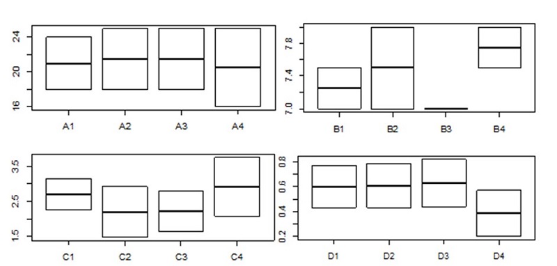 Graph 1. Variation of the 4 parameters measured in the first trial for immature larvae (L2). (A; length, B; maximum width, C; minimum width and D; weight), during the different stages of the process (1; before process, 2; post-dehydration, 3; post-impregnation and 4; post-curing). Y axis represents millimeters or grams for each variable. Box plot shows median, interquartile range (subdivided box) and standard deviation (outside lines) of variables. |
 Graph 2. Variation of 4 measures in the first trial for mature larvae (L3). (A; length, B; maximum width, C; minimum width and D; weight), during the different stages of process (1; before process, 2; post-dehydration, 3; post-impregnation and 4; post-curing). Y axis represents millimeters or grams for each variable. Box plot shows median, interquartile range (subdivided box) and standard deviation (outside lines) of variables. |
There is evidence of unequal behavior between the L2 and L3 larvae in their maximum and minimum widths. After several ascending and descending oscillations, both L2 and L3 larvae increased their maximum width at the end of the process. The same happened for L2 with minimum width, whereas L3 larvae decreased for this parameter. With respect to length and weight, a progressive decrease of these parameters occurred during all stages of the plastination process.
None of the p-values analyzed by the statistical study was significant for Oestrus ovis immature larvae (length, maximum width, minimum width and weight); weight changes in mature larvae were significant (p-value < 0.05).
|
LARVA
|
INITIAL (FIXATION- DEHYDRATION) |
DEHYDRATION -IMPREGNATION | IMPREGNATION - CURING | TOTAL (Initiation-End) |
P-VALOR (Initiation-End) |
|
| LENGTH | L2 | +2.38% | 0% | -4.65% | -2.38% | 0.577 |
| L3 | -3.02% | -0.28% | -4.26% | -7.42% | 0.106 | |
| MAXIMUM WIDTH |
L2 | +3.45% | +2.44% | +10.71% | +6.90% | 0.346 |
| L3 | -4.65% | +2.44% | +6.51% | +4.03% | 0.247 | |
| MINIMUM WIDTH |
L2 | -18.67% | +1.36% | +31.17% | +8.13% | 0.346 |
| L3 | +5.54% | -8.82% | -13.23% | -16.5% | 0.078 | |
| WEIGHT | L2 | +1.67% | +3.28% | -38.89% | -68.01% | 0.095 |
| L3 | -7.62% | -10.31% | -38.85% | -49.33% | 0.014* |
Table 2: Mean variation (L2 and L3) of morphometric parameters for each phase and the total plastination process in Oestrus ovis during the 1st test (results expressed in % on the initial value) and statistical significance (*): Significant values (p <0.05).
Second assay
Plastination protocol: During this test, 95% acetone was employed in the first bath (-20° C) in order to avoid shrinkage of the larvae. Table 1 shows comparative times for the different stages for the first and second tests. Impregnation was prolonged three times longer than in the first assay to ensure complete exchange of acetone by the reaction mixture. The times employed in each step were adapted to the needs of each assay, using objective measures for them (purity of acetone in dehydration, bubbling in impregnation, and excess of silicone in curing).

Figures 4a and 4b
Morphological results: On the second day of curing, one of the specimens collapsed, with an excessive and rapid exit of silicone from the surface. This situation gave the appearance of an adherent silicone layer that was difficult to eliminate from the ventral surface. At the end of processing, good morphological results were obtained in 9 of the 10 plastinated larvae (Fig. 4). Slight macroscopic variations can be observed in the plastinated individuals, but much less pronounced than those detected in the specimens of the previous test.
Histological images from the first trial (Fig. 3c, d) showed the thick cuticle of the larvae and the presence of a high number of enucleated cells in both plastinated and unplastinated parasites, in contrast with the second trial (Fig. 3e, f). This characteristic seems to be a consequence of the thick cuticle already mentioned above. A potential delayed entry of the formaldehyde during fixation could cause a process of cellular lysis that had an impact during all processes. To avoid this defect, larvae should be fixed with an incision in their cuticle.
Morphometric results: Values of the variables measured (expressed as a percentage) are shown in Table 3.
|
LARVA
|
INITIAL (FIXATION- DEHYDRATION) |
DEHYDRATION-IMPREGNATION | IMPREGNATION - CURING | TOTAL (Initiation-End) |
P-VALOR (Initiation-End) |
|
| LENGTH | L2 | +1.15% | -3.41% | -4.71% | -6.90% | 0.174 |
| L3 | +0.95% | +1.42% | -0.93% | +1.43% | 0.789 | |
| MAXIMUM WIDTH |
L2 | +3.68% | -8.47% | 0% | -5.10% | 0.181 |
| L3 | -1.61% | -0.70% | +1.18% | -1.15% | 0.187 | |
| MINIMUM WIDTH |
L2 | +2.01% | -1.38% | -3.90% | -3.32% | 0.0625 |
| L3 | +0.31% | -2.55% | -1.04% | -3.56% | 0.187 | |
| WEIGHT | L2 | -21.25% | +15.87% | -30.14% | -36.25% | 0.058 |
| L3 | -22.84% | +24.80% | -16.99% | -20.06% | 0.062 |
Table 3. Mean variation for each phase and the total plastination process in Oestrus ovis during the 2nd test (results expressed in % on the initial value) and statistical significance (*): significant values (p <0.05).
An overall decrease in all the parameters, except for the length in L3, were observed for both types of larvae in all phases of the plastination process. Weight decreased after the dehydration of the larvae, and increased after forced impregnation. The difference between results obtained for L2 and L3 before and after being plastinated were not significant, which shows that these plastinated specimens retained their morphology in an acceptable manner, in relation to their initial state (Graphs 3 and 4).
 Graph 3. Variation of 4 measurements in the second trial for immature larvae. (1 large, 2 max length, min length and 4. weight), during the different stages of the process (A; before process, B; post-dehydration, C; post-impregnation and D; post-curing). Y axis represents millimeters or grams for each variable. Box plot shows median, interquartile range (subdivided box) and standard deviation (outside lines) of variables. |
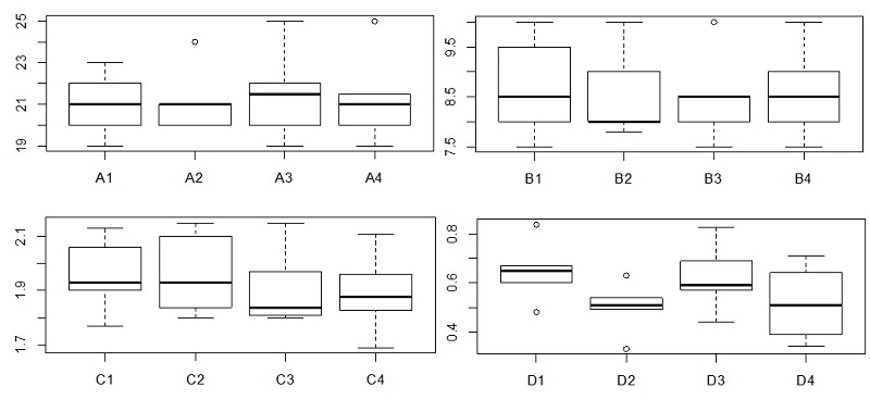 Graph 4. Variation of 4 measurements in the second trial for mature larvae (1 large, 2 max length, min length and 4. weight), during the different stages of the process (A; before process, B; post-dehydration, C; post-impregnation and D; post-curing. Y axis represents millimeters or grams for each variable. Box plot shows median, interquartile range (subdivided box) and standard deviation (outside lines) of variables. |
Plastination is a technique of conservation that produces samples of excellent quality and ease of handling, in contrast to the original biological specimens. In the present study, we have analyzed the possible use of plastination in parasitology, a discipline that has hitherto been largely unexplored.
For this objective, we started by employing the standard S10 protocol, adapting the technique by adjusting the times necessary for each phase. It has been possible to obtain correctly plastinated specimens, with no significant morphometric differences from their previous, formalin-fixed state, which are, therefore, very suitable for use in teaching or research. In addition, throughout the process, the times required to complete each phase have been reduced, compared with data published in previous trials, which would mean reductions in production time, and savings in cost of production (Kocevski et al., 2010).
Direct application of the standard S10 protocol did not enable us to produce an adequate final product. In Oestrus ovis larvae, the thickness of the cuticle may have prevented the circulation of fluids during the fixation, dehydration, and impregnation stages. The different level of development of cuticle in immature and mature larvae could be an element which may influence about the collapse of structures, so it seems appropriate to study each larval stage separately. This potential barrier during fixation could be the reason for enucleated cells, as a result of late penetration of formaldehyde, leading to cellular apoptosis. This fact could also be the cause of inadequate penetration of acetone and silicone during the dehydration and impregnation stages, causing a posterior collapse of the whole parasite during curing, favored by the internal spaces (coelom) that are present in the anatomy of these organisms. For this reason, incision of the cuticle is essential for correct plastination of these parasites. In such cases as these, it will be necessary to develop variations in the standard techniques in order to obtain satisfactory results, as has occurred in other studies, to solve the problem of a cuticle that interferes with the exchange of acetone and polymers, in fetuses or reptiles, among others (Ekim et al., 2017; Tiwari et al., 2012). The decreased time of the dehydration process in the second assay was related to the ventral incision of the larvae, because of the ensuing greater and faster contact between acetone and parasite. The increase in the available surface allows greater contact with silicone, increasing the period of impregnation. Curing time was reduced, to avoid the rigidity of specimens caused by exposure to crosslinker. Morphometric variations found during the second assay were less pronounced than in the first trial, indicating that this was a better protocol for Oestrus ovis larvae.
In conclusion, the morphology and morphometry of Oestrus ovis larvae, stages L2 and L3, can be preserved with the conventional S10 plastination protocol, with the addition of a previous incision in the cuticle. In general, more studies are necessary to establish the changes in the standard S10 protocol necessary to carry out effective plastination of different parasite specimens, and to evaluate their anatomical structures for the best results.
Asadi MH, Mahmodzadeh A. 2004: Ascaris plastination using S10 technique. J Int Soc Plastination 20: 24.
https://doi.org/10.56507/EDVF4592
DeJong K, Henry RW. 2007: Silicone plastination of biological tissue: cold-temperature technique - BiodurTM S10/S15 technique and products. J Int Soc Plastination 22: 2-14
https://doi.org/10.56507/ZLMJ7068
Ekim O, Merih Haziroglu R, Insal B, Bakici C, Akgün RO, Tunali S. 2017: A modified S10B silicone plastination method for preparation and preservation of scaled reptile specimens. Ankara Üniv Vet Fak Derg 64: 155-160.
https://doi.org/10.1501/Vetfak_0000002792
Essa I, Azzal G, Al-Azizz S, Abdulkhalek Sawad A, Samar GH. 2014: Plastination of arthropods using S10 technique. Basrah J Vet Res 11: 18-25.
Grondin G, Grondin GG, Talbot BG. 1994: A study of criteria permitting the use of plastinated specimens for light and electron microscopy. Biotech Histochem 69: 219-34.
https://doi.org/10.3109/10520299409106291
Kocevski Z, Stefanovska J, Ilieski V, Pendovski L, Atanaskova E. 2010: Improved determination of macroscopic parasite preparations using S10 modified plastination procedure. Macedonian Veterinary Review 2: 7-14.
Latorre RM, García-Sanz MP, Moreno M, Hernández F, Gil F, López O, Ayala MD, Ramirez G, Vázquez JM. Arencibia A, Henry RW. 2007: How useful is plastination in learning anatomy? J Vet Med Educ 34: 172-6.
https://doi.org/10.3138/jvme.34.2.172
McLaughlin JK. Formaldehyde and cancer: a critical review. 1994: Int Arch Occup Environ Health 66: 295-301.
https://doi.org/10.1007/BF00378361
Swenberg JA, Moeller BC, Lu K, Rager JE, Fry R, Starr TB. 2013: Formaldehyde carcinogenicity research: 30 years and counting for mode of action, epidemiology, and cancer risk assessment. Toxicol Pathol 41: 181-189.
https://doi.org/10.1177/0192623312466459
Tiwari S, Nandlal B, Shama Sundar NM. 2012: Plastinated fetus: 3D CT scan (VRT) evaluation. Indian J Dent Res 23: 686-688.
https://doi.org/10.4103/0970-9290.107411
Von Hagens G, Tiedemann K, Kriz W. 1987: The current potential of plastination. Anat Embryol 175: 411-21.
https://doi.org/10.1007/BF00309677