Department of Anatomy and Embryology, University of Nijmegen, The NETHERLANDS.
Modern imaging techniques require a basic knowledge of cross-sectional anatomy of the human body. In the present study, the technique of sheet plastination, introduced by von Hagens, was used for the 4 mm anatomical slices of a formalin-fixed human head. In the process of plastination water and lipids are replaced by polymers, which is subsequently hardened, resulting in dry, odourless and durable specimens. The P 35 plastination technique gives excellent differentiation between grey and white matter of the brain and offers excellent reference material for pre- and postdoctoral training in cross-sectional anatomy.
P35; cross sections;
R.T. de Boer-van Huizen, Department of Anatomy and Embryology, University of Nijmegen, The NETHERLANDS.

![]()


The wide application in the clinical neurosciences of imaging techniques such as computed tomography (CT) and magnetic resonance imaging (MRI) has aroused a growing interest in the use of anatomical sections of the human head and central nervous system (CNS). Excellent publications on sectional anatomy of the human head and CNS (Eycleshymer and Schoemaker, 1911; Koritke and Sick, 1983; Schnitzlein et al., 1983; de Groot, 1984; Kretschmann and Weinrich, 1984; Daniels et al., 1987; Gerhardt and Frommhold, 1988; Nieuwenhuys et al., 1988; Talairach and Tournoux, 1988; von Hagens et al., 1990) are available as anatomical guides for the analysis of CT and MRI sections. Study of actual sections of the human head would be preferable, particularly for teaching purposes. Macrosections of the human head or brain have some disadvantages in that they have to be kept in formalin, fall apart because of handling, and the differentiation between white and grey matter of the brain becomes less distinct in time due to the bleaching effect of the fixative. Cryomicrotomy (Rauschning et al., 1983) is a costly alternative requiring expensive equipment. Plastination, a technique of tissue preservation, introduced by von Hagens (1987), offers the possibility to prepare clean, dry, and easy to handle slices. In the process of plastination, water and lipids are replaced by polymers which are subsequently hardened resulting in dry, odourless and durable specimens. The polyester resin P 35 can be used for the production of opaque brain slices, which gives excellent differentiation between grey and white matter (von Hagens et al., 1987, 1990). In the present study, the P 35 plastination technique has been applied to 4 mm anatomical slices of a formalin-fixed human head.
The standard P 35 technique according to von Hagens (1985, 1989; von Hagens et al., 1987) was used. This technique involves the following steps:
SPECIMEN PREPARATION FOR FIXATION AND SAWING:
Our first "plastination experiments used the head of a human, male, cadaver (73 years of age) which had been preserved in embalming fluid for over three years. The embalming fluid consisted of 500 ml ethanol 96%, 1000 ml formalin 40%, 25 ml phenol liquefied 80%, 300 gm sodium chloride, 300 gm chloral hydrate and 300 ml glycerin in 10 liters of water. The head was removed from the body and rinsed in running tap water for two days. After rinsing, it was placed in a styrofoam box filled with water and frozen at - 20° C with the head oriented so that slices could be sawn parallel to Talairach's (1952) bicommissural (ACPC: anterior commissure - posterior commissure) plane (Bergvall et al., 1988). After three days in the deep freezer, the frozen styrofoam box, ice, and head were sawn using a handsaw into 4 mm thick slices. The macrosections of the head were placed on glass-plates. After defrosting, the slices were gently rinsed with running tap water to remove the sawdust and pieces of the styrofoam box. After making color photographs of the slices, the slice and plate were submerged into 4% formalin where they were kept until further processing.
DEHYDRATION:
The 4 mm thick head slices were carefully put between stainless steel grids and rinsed for 8 hours in running tap water to remove the fixative. To prevent the brain from becoming too brittle after dehydration in cold acetone, the slices were placed into a solution of 4% sucrose in distilled water at 4"C over night. According to von Hagens1 protocol (1987), dehydration was performed by freeze substitution using cold acetone of -25 °C. The acetone was changed twice and the specimens were kept in the cold acetone for 2 - 14 days.
IMPREGNATION:
From the cold acetone, the specimens were quickly immersed in a polymer-mix [20 ml of harder (A9) to one liter P 35] at 4°C. After gently removing excessive air-bubbles with the aid of low (minimal) vacuum for 20 - 30 minutes, the specimens were left over night in the first polymer-mix at 4°C. The polymer-mix was changed three times at 24 hour intervals and low vacuum was used for removing air- bubbles each time. Forced impregnation was started after the third change of polymer-mix as the vacuum was increased to 12 mm Hg over a 4 - 5 hour period of time. Vacuum was maintained at 12 mm Hg over night in the dark vacuum chamber at room temperature.
PREPARATION OF THE PLASTINATED SHEETS:
The head slices were carefully removed from the stainless steel grids and placed into the flat chamber consisting of four glass-plates. The two inner plates were separated by a flexible, elastic gasket. A detailed description of this technique is found in the "Heidelberg Plastination Folder" (von Hagens, 1985; von Hagens et al., 1987). To construct the flat chamber for the 4 mm thick slices, a 6 mm thick gasket was used. A small nylon string was connected to the skull to prevent the slice from sinking to the bottom of the flat chamber. After filling the flat chamber with freshly prepared polymer- mix it was again placed into the vacuum chamber and kept under low vacuum for several (3-5) hours to remove the air bubbles from around the specimen, and even more important, most of the small fat globules. After all air bubbles and most of the fat globules had floated to the top of the polymer, the top of the flat chamber was closed utilizing the ends of the gasket. Polymerization was initiated with a 200 Watt UV-A light source for 45 minutes on both sides. After light curing, the flat chamber was kept in an oven at 40 °C for six days to complete the process of polymerization. When this process was completed, the flat chamber was dismantled. The plastinated slice was covered with plastic foil, excessive polymer was trimmed away, and the specimen was polished with waterproof sandpaper and abrasive cloth.
ACETONE VAPOR MONITORING:
Acetone vapor levels were monitored at various stages of the dehydration process. A hexane calibrated TLV-Sniffer was used to measure acetone vapor levels in the deep freezer and at the inhalation level during manipulation of the specimens on the grids and while pouring acetone from one container to another. Vapor levels from pouring both cold (- 25 °C) and warm acetone (20 °C) were measured and recorded.
The contrast between grey and white matter of the brain is enhanced during the process of plastination as noted by the 4 mm thick macrosection of the human head (preplastination) and the resulting plastinated slice (Figs. 1, 2). Within the thalamus, the various nuclei can be discerned. Furthermore, large fiber bundles such as the radiatio optica and the forceps major (posterior) of the corpus callosum can easily be identified. The shrinkage of the brain and other soft tissues was minimal. Figure 3 shows a plastinated slice passing through the orbit, the external acoustic meatus, the inner ear and the fossa cranii posterior. The dentate nucleus and pontine nuclei stand out in the surrounding white substance of the cerebellum and pons, respectively. By examining the plastinated slices under a dissecting microscope or with a magnifying-glass small structures such as nerves, blood vessels and meninges can be studied in detail (Fig. 4).
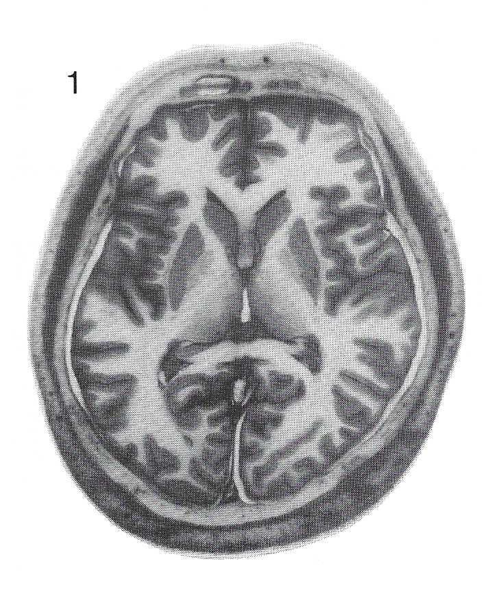 Figure 1. Section of the human head, parallel to Talairach's (1952) bicommissural plane. 4 mm thick, unstained, X 0.47. |
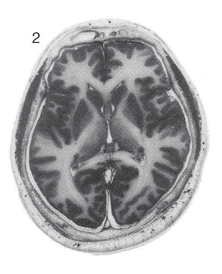 Figure 2. The resulting plastinated slice (same slice as in figure 1), processed by P 35 sheet plastination. 4 mm thick, unstained, X 0.47. |
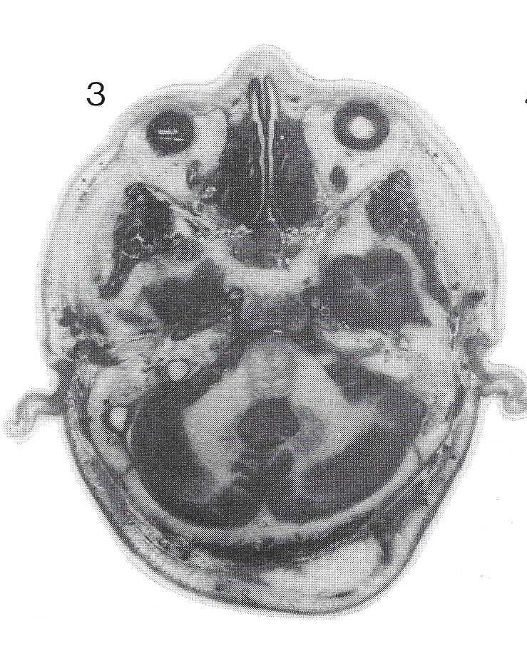 Figure 3. Plastinated slice of the human head. 4 mm thick, unstained, X 0.47. |
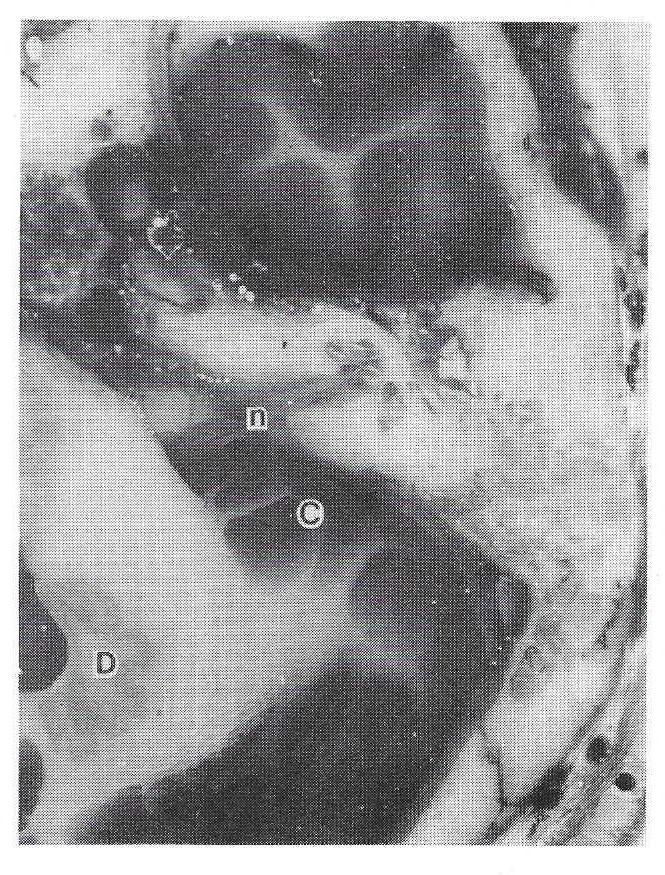 Figure 4. A close-up (higher magnification) of the sheet shown in figure 3 revealing structures at the cerebellopontine angle and the internal ear. Cerebellum (Ce), Dentate nucleus (De), Cranial nerve eight (n VIII). 4 mm thick, unstained, X 1.25. |
The section in Figure 3 shows the disturbing effects of lipids. The fat globules may accumulate on the surface of the slice.
Acetone concentrations above the open dehydration tank while specimens were being manipulated was 5,000 ppm; while at the inhalation level, the concentration was 850 ppm. When cold acetone (-25 °C) was poured into a dehydration vat, the acetone vapor level at the inhalation level was 45 ppm. When 20°C acetone was similarly poured into the vat, the vapor level at the inhalation zone was 450 ppm.
To aid our teaching program, sheet plastination of slices of the human head has been utilized in our laboratory. As already known, the P 35 technique gives excellent differentiation between grey and white matter of the brain (von Hagens et al., 1987, 1990). Macrosections of the head allow the study of the brain and its surrounding structures, i.e., the brain "in situ". Although the process of plastination is time-consuming, the availability of dry, odourless and durable specimens of the human head provides reference material for pre- and postdoctoral training in crosssectional anatomy which is convenient to handle.
Specimens were dehydrated for 2 - 14 days, however, in keeping with von Hagens' data (1989), 4 days appeared to be enough time for adequate dehydration. Longer periods of dehydration can result in extraction of too much lipid from the brain tissue.
The presence of fat globules on the surface of a plastinated slice is most likely due to the extraction of f at f rom the bone marrow or from an atheromatous cyst in the neck region. It should be noted that during dehydration in acetone and impregnation in the polymer some lipids are extracted from the tissue. For brain tissue, the extraction of lipids should be limited as the differentiation between grey and white matter is achieved by preservation of lipids (von Hagens et al., 1987). Since the skull and skin are still part of the specimen, the lipids from these tissues may cause these disturbing artifacts. They appeared as little white fat globules that sometimes cover the whole plastinated brain slice. These artifacts can be removed using a nylon brush and xylene to dissolve and remove the fat globules. As a result, the surface of the plastinated sheet will be covered with small depressions. These can be eliminated by placing the plastinated slice back into a 1:1 polymer/acetone mixture, for one or two days and then back in the flat chamber where the sheet plastination process is repeated. However, an 8 mm gasket is used to replastinate.
Since dehydration by freeze substitution (cold acetone) is recommended, an inherent problem with the plastination technique is acetone safety. A mixture of acetone and air may be highly explosive.
Measurements of the concentration of acetone vapors during our plastination work (Plas, 1991), showed that the highest concentration of acetone was found in the deep freezer while manipulating the specimens on the stainless steel grid in one of the open acetone tanks. This concentration was 5000 ppm just above the open tank. At the inhalation level during this maneuver, the highest concentration of acetone measured was 850 ppm. During the step in which cold acetone of -25 °C was poured out of the tank during the changing of acetone, the highest concentration at the level of inhalation was 45 ppm. When 20 °C acetone was poured, the concentration was 450 ppm. The lowest explosion level for acetone vapors is 2.3% (23,000 ppm). Our highest concentration of acetone in the deep freezer was well below this point. Therefore, the risk for explosion did not exist. The use of an explosion-safe deep freezer, however, has to be emphasized. It should also be stressed that for inhalation these concentrations of acetone are high, so it is advisable to work under a fume hood and if this is not possible, one should wear a mask. As the components of the P 35 polymer are not published, to measure their concentration was not possible. However, the vapor pressure of P 35 is very low. We recommend to work as clean as possible and wear protective clothing, mask and gloves.
Our next step in plastination will be to make other series of plastinated sections in frontal and sagittal planes.
ACKNOWLEDGEMENTS
The authors wish to express their gratitude to Dr. A.H.M. Lohman of the Department of Anatomy of the Free University of Amsterdam, for offering the opportunity to use their vacuum chamber; Mr. H.J.M. Remers for skillfully preparing the frozen sections of the head; Mr. T.W. Spaan for giving the finishing touch to the plastinated slices; and the collaborators of the Department of Photography of the University of Nijmegen for assistance with photography.
Bergvall U, C Rumeau, Y van Bunnen, JM Corbaz, M Morel: External references of the bicommissural plane. In: Brain Anatom y and Magnetic Resonance Imaging (A. Gouaze, G. Salamon, eds). Berlin-Heidelberg, Springer-Verlag, pp 2-10, 1988.
https://doi.org/10.1007/978-3-642-72709-2_2
Daniels DL, VM Haughton, TP Naidich: Cranial and Spinal Magnetic Resonance Imaging. Raven Press, New York, 1987.
https://doi.org/10.1097/00002142-198812000-00010
De Groot J: Correlative neuroanatomy of computed tomography and magnetic resonance imaging. Lea & Febiger, Philadelphia, 1984.
Eycleshymer AC, DM Schoemaker: A Cross- section Anatomy. Appleton, New York-London, 1911 (reprint 1970: Butterworths, London).
Gerhardt P, W Frommhold: Atlas of Anatomic Correlation in CT and MRI. Thieme Verlag, Stuttgart, 1988.
Koritke JG, H Sick: Atlas of Sectional Human Anatomy, Vol.1: Head, Neck, Thorax. Baltimore, Munchen, Urban & Schwarzenberg, 1983.
Kretschmann HJ, W Weinrich: Neuroanatomie der Kraniellen Computer Tomography. Thieme Verlag, Stuttgart, 1984.
Nieuwenhuys R, J Voogd, C van Huyzen: The Human Central Nervous System. A synopsis and atlas. Third revised edition, Berlin-Heidelberg, Springer-Verlag, 1988.
https://doi.org/10.1007/978-3-662-10343-2
Plas AM: Arbeidshygienische metingen inbeddingsproces. Internal report Health and Safety Service University of Nijmegen, 1991.
Rauschning W, K Bergstrom, P Pech: Correlative craniospinal anatomy studies by computed tomography and cryomicrotomy. J Comp Ass Tomography 7:9-13, 1983.
https://doi.org/10.1097/00004728-198302000-00002
Schnitzlein HN, EW Harley, FR Murtagh, L Grendley, JT Fargher: Computed Tomography of the Head and Spine. Urban & Scharzenberg, Baltimore, Munich, 1983.
Talairach J, J de Ajuriaguerra, M David: Etudes ste're'otaxiques des structures enc6phaliques chez I'homme. Presse Med 28:605-609, 1952.
Talairach J, P Tournoux: Co-planar Stereotaxic Atlas of the Human Brain. Thieme Verlag, Stuttgart, 1988.
von Hagens G: Heidelberg Plastination Folder. Anatomisches Institut 1, Universita't Heidelberg, D-6900 Heidelberg, 1985.
von Hagens G: Die Scheibenplastination. Internal report. Anatomisches Institut 1, Universita't Heidelberg, 1989.
von Hagens G, K Tiedemann, W Kriz: The current potential of plastination. Anat Embryol 175:411- 421, 1987.
https://doi.org/10.1007/BF00309677
von Hagens G, A Whalley, R Maschke, W Kriz: Schnittanatomie des menschlichen Gehirns. Ein photographischer Atlas plastinierter Serienschnitte. Steinkopff Verlag, Darmstadt, 1990.
https://doi.org/10.1007/978-3-642-93670-8