Departement de Chimie-Biologie, Universite du Quebec a Trots-Rivieres, Trois-Rivieres, Quebec, Canada
Vascular injections of colored mixtures were probably not performed before the seventeenth century. They are intended to allow a more detailed description of arteries, veins and lymph vessels, but also to produce specimens to be exhibited in anatomical and natural sciences museums, usually after corrosion. This paper gives a summary of the development of vascular injection procedures and mixtures in the seventeenth and eighteenth centuries, with special reference to the injection of the heart vessels (coronary arteries, venae cordis minimae, and lymphatics).
Vascular injections, Coronary arteries, Venae cordis minimae, Lymphatics of the heart
Dr. R. Olry, Departement de Chimie-Biologie, Universite du Quebec a Trois-Rivieres, C.P. 500, Trois-Rivieres, Quebec, Canada G9A 5H7. Telephone: 819 376 5053 / Fax: 819 376 5084 Email: Regis_Olry@uqtr.uquebec.ca

![]()


The knowledge of the course and ramifications of blood vessels was based for a long time on the dissection of non injected vascular systems. Only large vessels could there- fore be described, for the small branches were usually cut off during the dissection. Galen recommended introducing a wooden probe into the vessels during the
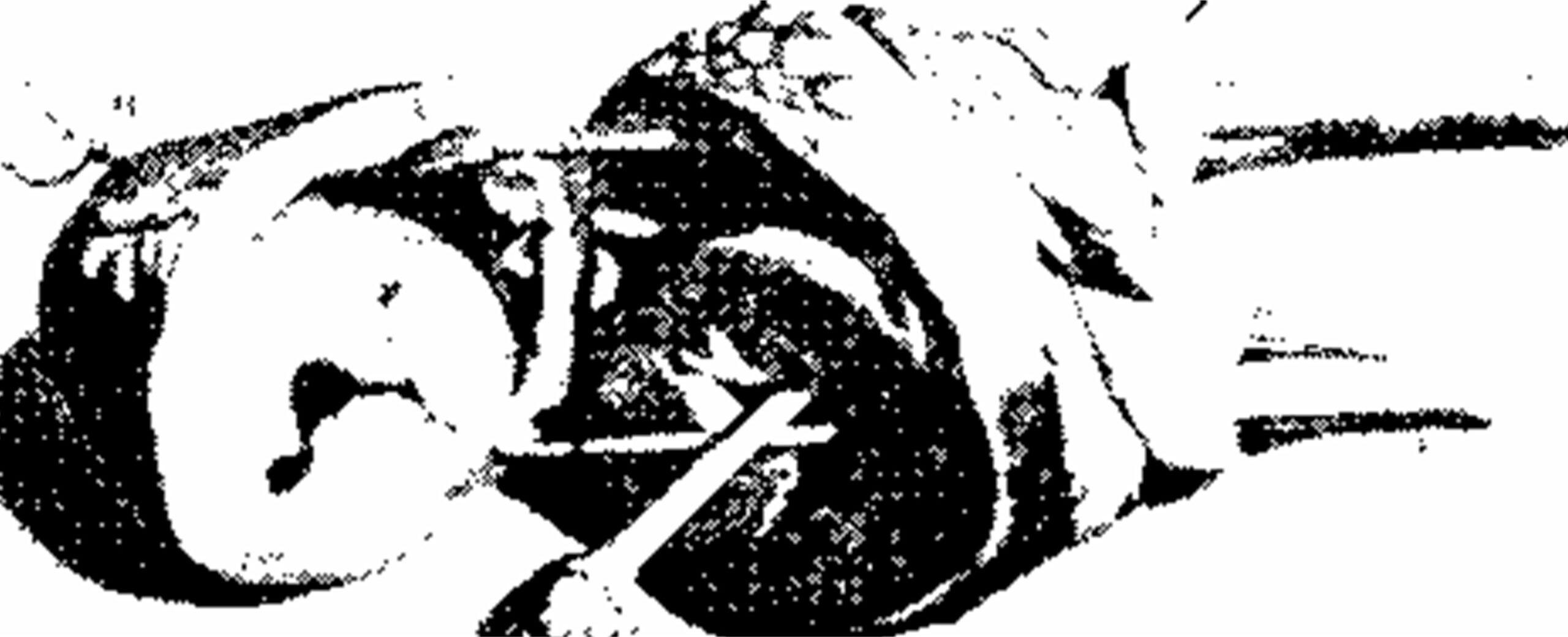
Figure 1. Plate 9 of Bidloo's treatise, engraved by Gerard de Lairesse (1685). Wooden probes are introduced into the heart cavities to show their communications with the large vessels.
dissection, so that the knife could not damage their walls. This procedure was still in use in the seventeenth century. Covert Bidloo (1649-1713), among others, depicted wooden probes in the heart cavities on a plate of his
famous 1685 treatise (figure 1). The problems with these probes were two in number. First, they could not be introduced into very small vessels. Second they were not flexible and therefore compromised the course and relationships of the vascular system. That is why it became necessary to develop vascular injection procedures on the one hand, and to find color injections mixtures on the other hand (table 1). In addition to the scientific aspect of these procedures (detailed anatomical description of the vascular system), vascular injections were also intended to pro- duce long lasting specimens for anatomical and natural sciences museums which drew crowds in the eighteenth century.
| Author | Date | Substance | Equipment |
| A. Gigliani (?) | Early XlVth century | Various colored substance | 7 |
| L. da Vinci (?) | 1504-1507 | Wax | 7 |
| G. B. da Carpi | 1522 | Warm water | Syringe |
| A. Vesale | 1543 | 7 | Siphon |
| W. F. von Hilden | 1615 | 7 | Cannula linked to a bladder |
| J. Swammerdam | 1672 | Melted wax | Copper syringe |
| G. Homberg | 1699 | Lead, tin, bismuth | Pneumatic apparatus |
| R. Vieussens | 1706 | Saffron tinting | ? |
| A. C. Thebesius | 1708 | Water, colored wax | ? |
| F. Ruysch | 1726 | "Materia ceracea" | 7 |
| Ronhaut | 1718 | Gelatin | ? |
| G . A. Langguth | 1746 | 7 | Sipho anatomicus |
The development of vascular injection procedures
Vascular injections may have been performed by anatomists as far back as in the Middle Ages. Alessandra Gigliani, who assisted the famous Bolognese anatomist Mondino dei Luzzi (ca. 1275-1326) in his lectures, is sometimes regarded as the first one to have been successful in injecting blood vessels with various colored substances» (Martin, 1934). However, no information could be found concerning the nature of these substances and the injection procedure. Moreover, this hypothesis has been rejected by most of historians of anatomy, who question Gigliani's biographical data (Wolf-Heidegger and Cetto, 1967; Olry, 1997).
Leonardo da Vinci (1452-1519) is known to have per- formed wax injections into the brain ventricles, and
tions of a solidifying liquid into the body» (Cazort et al., 1996). We could therefore infer that he probably also used vascular injections to study the course of blood vessels and the morphology of the heart cavities although there is no definitive proof that he used vascular injection (O'Malley and Saunders, 1952; Huard, 1968; Kurz, 1992).
Though Andre Vesale (1514-1564) described in his 1543 treatise a U-shaped tube (siphon) to perform injections, it seems that he never used it for the injection of blood vessels (Kurz, 1992). Moreover, this instrument appears neither on the plate depicting the dissection and preparation instruments in the 1543 edition, nor on the frontispiece of the fifth 1604 edition. A detailed description of this «Sipho anatomicus» was made about two centuries later (1746) by the German anatomist Georg August Langguth (1711-1782) (figure 2).
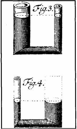 Figure 2. The «sipho anatomicus» by G. A. Langguth (1746). |
 Figure 3. The injection apparatus described by W. F. von Hilden (1646). A and B: tube linked to a cannula. C: dried bladder. D: funnel. E and F: faucets. |
In the mid-seventeenth century, the surgeon Wilhelm Fabricius von Hilden (1560-1634) used an injection apparatus composed of a cannula linked to a dried bladder. A funnel and two faucets allowed the filling of the bladder and prevented the back flow of the injected mixture from the vessels to the cannula (figure 3). Subsequently, many famous anatomists tried to improve on this kind of injection appara- tus: Regnier De Graaf (1668), Francis Glisson (1677), Rich- and Lower (1708), Stephen Hales (1733-1734), Alexander Monro (1733), Johann Nathanael Lieberkuhn (1789) (figure 4), Ludwig Teichmann (1879), and Gustav Schmiedel (1930), among others.
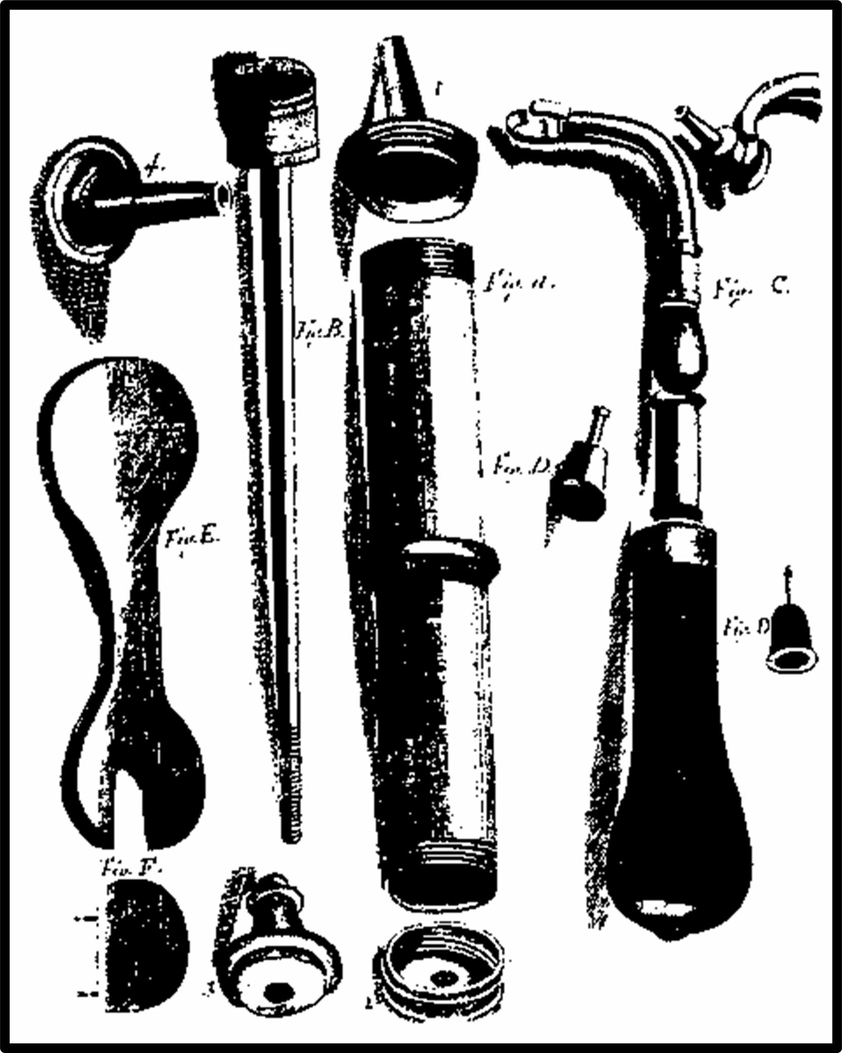
Figure 4. Syringes for vascular injections after J. N. Lieberkuhn (1789).
The development of vascular injection substances
Many experiments were performed using various vascular injection mixtures in order to enhance the results (table 1). As far back as in 1522, Giacomo Berengario da Carpi (ca. 1460-1530) performed vascular injections with warm water, using a syringe. Thirty years later, Bartolomeo Eustachi (1520-1574) used the same procedure on renal arteries, and could observe the filling of the bladder.
However, the best vascular injection procedure results were obtained by the Dutch anatomists Jan Swammerdam (1637-1680) and Frederik Ruysch (1638-1731). In 1672, Jan Swammerdam injected melted wax into the vessels of the uterus using a copper syringe (Fort, 1902). The specimens were so attractive that Frederik Ruysch decided to improve on the method. In the early eighteenth century, he was acknowledged as the «apostle of the injection technique* (Hagelin, 1989), and his striking specimens drew crowds in anatomical museums. Fontenelle said that «in a way, Ruysch's mummies prolong life, whereas Egyptian mummies only prolong death» (cited by Fort, 1902). The vascular injections of the specimens were so successful that Ruysch wanted to keep the composition of his injection mixture secret. He only called it «Materia ceracea». However, its composition was published twelve years after his death. It was composed of tallow, white wax, cinnabar, and «sometimes other substances, depending on the seasons* (Kurz, 1992).
In the late seventeenth century, Homberg injected blood vessels with a mixture of lead, tin and bismuth (1699); un- fortunately, the results were disappointing. Finally, gelatin was used for the first time in vascular injection by Ronhaut, the surgeon to the King of Sardinia (1718).
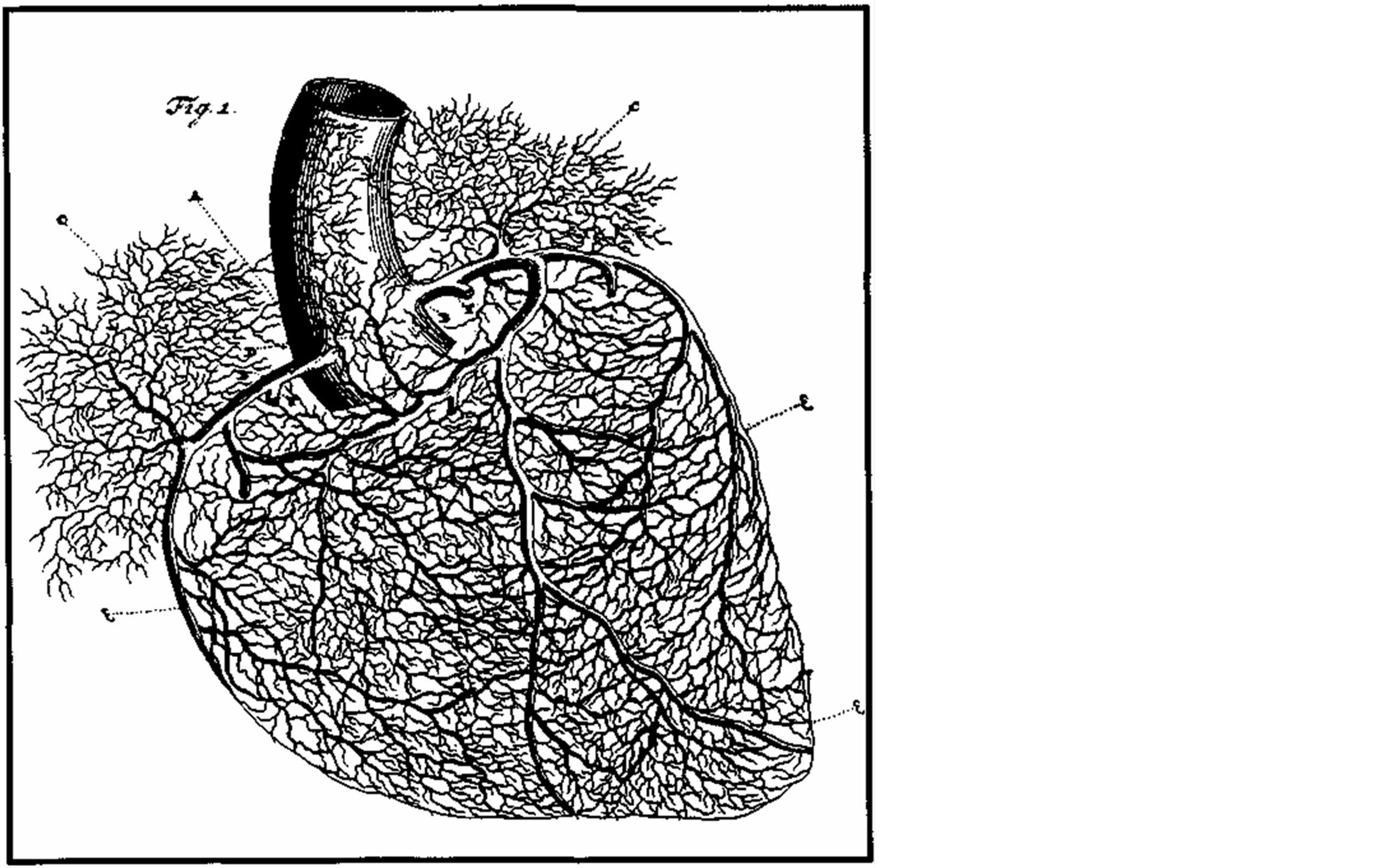
Figure 5. Vascular injected specimen of the heart by F. Ruysch (1726).
A wonderful specimen of injected coronary arteries was depicted in Frederik Ruysch's Opera omnia (1726) (figure 5). This specimen shows the ascending aorta, the right coronary artery and some of its ventricular branches, and the left coronary artery with its division into anterior interventricular and circumflex arteries. Numerous atrial rami are to be seen, arising from both coronary arteries. A large left conus artery is depicted, leaving the anterior interventricular artery near its start, and anastomosing on the conus with that of the right coronary artery.
The discovery of the opening of the venae cordis minimae into all cardiac cavities, is commonly attributed to the German anatomist Adam Christian Thebesius (1708). However, the existence of these minimal veins (Aho, 1950), which are more difficult to demonstrate than larger cardiac vessels, was first reported by Raymond Vieussens in a letter addressed to M. Boudin in 1706. Raphael Bienvenu Sabatier gives a detailed account of Vieussens' procedure in a memoir of 1792. For this experiment, Vieussens used two human hearts. He tied both venae cavae, the pulmonary trunk and the aorta. Then, he injected slowly a saffron tinting into the left coronary artery, and could observe that both atrial and ventricular left cavities became full of yellowish liquid. Moreover, the right cavities remained uncolored. Thebesius only confirmed the existence of these veins in his Latin dissertation (1708), by injecting water and colored wax into the cardiac veins.
Though Olof Rudbeck (1630-1702) discovered the subpericardial lymph vessels as far back as 1653, the systematization of the lymphatics of the heart was not under- stood before the second half of the nineteenth century (for the history of injection procedures of lymphatics in general, see Olry and Motomiya, 1997). The subendocardial lymph network was discovered by Eberth and Belajeff (1866), the epicardial ventricular lymph network by Sappey (1874), and the epicardial atrial lymph network by Rainer (1907) and Mouchet (1909).
Aho A: On the venous network of the human heart and its arteriovenous anastomoses. Ann Med Exp Biol Fenn 29 (Suppl 1): 1-90, 1950.
Bidloo G: Anatomia humani corporis, Centum & quinque Tabulis, per artificioss. G. de Lairesse ad vivum delineatis, Demonstrata, Veterum Recentiorumque Inventis explicata plurimisque, hactenus non detectis, illustrata. Amstelodami: J. a Someren, 1685.
Cazort M, Kornell M, Roberts KB: L'ingenieuse machine humaine. Quatre siecles d'art et d'anatomic. Ottawa: Musee des Beaux-Arts du Canada, 1996.
da Carpi GB: Isagogae breves perlucide ac uberrime in anatomiam humani corporis, a communi medicorum academia usitatam, a Carpo... in lucem date... Bononiae: Hectoris, 1522.
de Graaf R: De virorum organis generationi inservientibus, de clysteribus, et de usu siphonis in anatomia. Lugduni Batavorum & Roterodami: Hack, 1668.
Eberth, Belajeff: Ueber die Lymphgefasse des Herzens. VirchowArch37: 124-130, 1866.
https://doi.org/10.1007/BF02116555
Fort JA: Histoire de 1'anatomic. In: Anatomic descriptive et dissection. Paris: Vigot Freres, 6eme edition, vol. 3, pp. 905-1029, 1902.
Glisson F: Tractatus de ventriculo et intestinus. Cui praemittur alius de partibus continentibus in genere; & in specie de iis abdominis. Londini: H. Brome, 1677.
Hagelin O: Rare and important medical books in the library of the Swedish Society of Medicine. Stockholm: Svenska Lakaresallskapet, 1989.
Hales S: Statical essays... containing vegetable staticks; or, an account of some statical experiments on the sap in vegetables... Also a specimen of an attempt to analyse the air... Containing haemastaticks; or, an account of some hydraulick and hydrostatical experiments made on the blood and blood-vessels of animals. London: Innys, 1733-1734.
Homberg G: Hist Acad, 1699.
Huard P: Leonard de Vinci. Dessins anatomiques (anatomic artistique, descriptive et fonctionnelle). Paris: R. Dacosta, 1968.
Kurz H: Die Darstellung von Blutgefassen und Hohlraumen. Zur geschichtlichen Entwicklung der Injektionstechnik. Basel: Anatomisches Institut der Universitat Basel, 1992.
Langguth GA: De siphonis anatomici usu parum anatomico commentatio... Adjecta est tabula aeriea. Witembergae, Lieberkiihn JN: Abhandlungen von anatomischen
Einspritzungen und Aufbewahrung anatomischer Praparate, 1789.
Lower R: Tractatus de corde. Item de motu & colore sanguinis et chyli in eum transitu. Editio quinta. Lugduni Batavorum, J. van der Linden, 1708.
Martin CR: Histoire de 1'enseignement de 1'anatomie. Lyon: Librairie Arnette, 1934.
Mouchet A: Les vaisseaux lymphatiques du coeur chez rhomme et quelques mammiferes. J Anat Physiol 45: 433-458, 1909.
Olry R: Homo dissectus. Petites histoires de grands anatomistes. Trois-Rivieres: Les Editions du Bien Pub- lic, 1997.
Olry R, Motomiya K: Paolo Mascagni, Ernest Alexandre Lauth and Marie Philibert Constant Sappey on the Dis- section and Injection of the Lymphatics. J Int Soc Plastination 12 (2): 4-7, 1997.
https://doi.org/10.56507/RXFY8836
O'Malley CD, Saunders JB de CM: Leonardo da Vinci on the human body: The Anatomical, Physiological and Embryological Drawings of Leonardo da Vinci. New York, 1952.
Rainer FJ: Ueber das Vorkommen von subepicardialen Lymphdrüsen beim Menschen. Anat Anz 31: 46-49, 1907.Ronhaut: cited by Fort (1902).
Rudbeck O: Nova exercitatio anatomica, exhibens ductus hepaticos aquosos, & vasa glandularum serosa, nunc primum inventa, aeneisque figuris delineata... Arosiae: Lauringerus, 1653.
Ruysch F: Opera omnia, 1726.
Sabatier RB: Sur les veines de Thebesius. In: Traite complet d'anatomic ou description de toutes les parties du corps humain. Paris: T. Barrois, vol. 4, pp. 388-405,1792.
Sappey MFC: Anatomie, physiologic, pathologic des vaisseaux lymphatiques considered chez l'homme et les vertebres. Paris: A. Delahaye & E. Lecrosnier, 1874.
Swammerdam J: Miraculum naturae, seu uteri muliebris fabrica, 1672.
Thebesius AC: Disputatio medica inauguralis de circulo sanguinis in corde. Lugduni Batavorum: A. Elzevier, 1708.
Vesale A: De humani corporis fabrica libri septem. Basileae: Oporinus, 1543.
Vesale A: Anatomia. Venetiis: I. A. et I. de Franciscis, 1604. Vieussens R: Nouvelles decouvertes. Lettre adresse"e a M.
Boudin Conseiller d'Etat, et premier Medecin de Monseigneur, 1706.
von Hilden WF: Opera observationum et curationum. 1646.
Wolf-Heidegger G, Cetto AM: Die anatomische Sektion in bildlicher Darstellung. Basel New York: S. Karger, 1967.