Zentrum der Morphologic J.W. Goethe-Universitat Frankfurt, West Germany
Teaching morphological aspects in neuroscience is accompanied by two main problems. 1. Wet brain tissue is inconvenient to handle and is easily destroyed when demonstrated a number of times. 2. Macroscopic demonstration of subcortical nuclei is difficult in unstained sections. In particular, subdivisions of nuclear greys are difficult to delineate. These two problems are accentuated when demonstrating human fetal brain tissue. These problems can be prevented by the methods described in this paper which combine the advantages of staining of brain tissue sections with those of plastination (Ulfig and Wuttke, 1990). An additional procedure, prior to staining, is necessary for the human fetal brain tissue because of its high vulnerability. Fetal brain tissue should be embedded in a suitable medium before cutting and staining, otherwise these sections will not withstand the staining procedure.
staining; human fetus; s10; plastination; silicone
Norbert Ulfig - Zentrum der Morphologic J.W. Goethe-Universitat Frankfurt, West Germany

![]()


Teaching morphological aspects in neuroscience is accompanied by two main problems. 1. Wet brain tissue is inconvenient to handle and is easily destroyed when demonstrated a number of times. 2. Macroscopic demonstration of subcortical nuclei is difficult in unstained sections. In particular, subdivisions of nuclear greys are difficult to delineate. These two problems are accentuated when demonstrating human fetal brain tissue. These problems can be prevented by the methods described in this paper which combine the advantages of staining of brain tissue sections with those of plastination (Ulfig and Wuttke, 1990). An additional procedure, prior to staining, is necessary for the human fetal brain tissue because of its high vulnerability. Fetal brain tissue should be embedded in a suitable medium before cutting and staining, otherwise these sections will not withstand the staining procedure.
ADULT HUMAN BRAIN TISSUE
Fixation: Fix human brain material by immersion in a 4% aqueous formaldehyde solution for several months.
Cutting: Rinse in running tap water overnight. Cut sections (1 to 4 mm) with the aid of a freezing microtome. [Warm a plane block of aluminum to about 35 ° C. Place this block to the surface of the frozen tissue for a few seconds (the time required is determined after a few trials). Sections should be cut at the junction of frozen and thawed tissue. Sections may also be cut with the aid of a microtome or a knife (0.5 to 2 cm].
Oxidation: Rinse sections in running tap-water and oxidize in freshly prepared performic acid [100 ml perhydrol (30% H2O2) to 900 ml 100% formic acid] for one hour. Sections must be kept in motion during oxidation. Handle the performic acid solution with great care under a chemical hood. Following one hour of oxidation, wash sections under tap-water for at least one hour and considerably longer when thick microtome sections are used. The previous color of the sections should have returned prior to staining.
Staining: Stain sections with either of two stains: 1) Astra blue [dissolve 0.1 g astra blue (Merck) in 1000 ml distilled water and add 1 ml HCI (37%)] for two days. Keep sections in motion, or 2) After rinsing in 70% ethanol for 15 minutes, stain with aldehvdefuchsin for two days. Keep sections in motion. Prepare aldehydefuchsin stock solution by dissolving 0.5 g pararosanilin (Chroma) in 100 ml 70% ethanol and then add 1 ml of 25% HCI and 1 ml 100% crotonaldehyde. Shake briefly and let react for one week. Use this stock solution for one week only. Prepare the staining solution by adding 6 ml stock solution, 100 ml distilled water, 50 ml 100% formic acid, and 5 ml performic acid (prepared as for oxidation) to 400 ml of 96% ethanol and filter.
Dehydrate: Sections for 12 hours in each ethanol solution (70, 80 and 96%), and then for at least 24 hours in 100% ethanol. In the 96 and 100% ethanol solutions, place the sections between filter paper and perforated porcelain plates.
Plastination procedure: Transfer the sections, covered by filter paper and between perforated plates, to 100% acetone for one day (1 - 4 mm sections) or three days (0.5 - 2 cm sections). Place the sections, between perforated porcelain plates, into the S10/S3 polymer mix in the vacuum chamber for three days (1 - 4 mm sections) or seven days (0.5 - 2 cm). Remove sections and drain excess silicone. For curing, transfer the sections to a curing chamber with a large volume of S6. During the first few hours of curing, excess silicone must be regularly wiped off to avoid superficial precipitations. After the surface is cured, transfer sections to an air-tight container, which has calcium chloride in it to remove the moisture. The sections are ready after two weeks.
FETAL HUMAN BRAIN TISSUE
Fixation: Fix by immersion in a 4% aqueous formaldehyde solution for several weeks.
Dehydrate: in a graded series of ethanol. After dehydration in 100% alcohol, place into a refrigerated mixture (1:1) of 100% diethyl-ether and absolute ethanol.
Embedding: with Cedukol (Merck, Darmstadt), produces optimal results. Soak in 2%, 4%, and 8% solutions of celloidin. Finally, concentrate to a 16% solution of celloidin. Harden in 70% ethanol (Romeis, 1989).
Cutting: Cut at 0.5 - 5 mm with the aid of a freezing microtome. The block must always be wetted by 70% alcohol and sections must be stored in 70% alcohol. Excess celloidin should be cut away.
Staining: Rinse the sections in distilled water for 10 minutes before the staining procedure. Stain section with Darrow-red free-floating (Powers et al., 1960; Powers and Clark, 1963 slightly modified). Prepare a stock solution: dissolve 0.1 g Darrow-red (Aldrich Chemical Company, Inc., USA) in 4.8 ml acetic acid and 395.2 ml distilled water. Stir and boil gently for 10 minutes. Cool to room temperature and filter. Use 4 parts of stock solution and 1 part 0.2 molar sodium acetate solution to prepare the staining solution which should be used the same day. Stain the sections for 12 hours. Keep sections in motion while staining.
Dehydration: Place sections into 70% ethanol for 20 minutes and observe carefully. If sections lose color, transfer them to 96% ethanol immediately. Place sections between filter paper and perforated porcelain plates to obtain flat sections. Leave sections in 96% ethanol for one hour and transfer into 100% propylalcohol for 60 minutes (between plates). Do not use 100% ethanol. Then dehydrate sections free-floating in 100% propylalcohol for 20 minutes. If sections are stained too intensively, keep sections in 100% propylalcohol until the desired intensity is achieved.
Plastination procedure: The same procedure as described above.
ADULT HUMAN BRAIN TISSUE
Both frozen sections, as well as, thicker macrotome sections may be used for these staining techniques. A series of the thinner frozen sections provides more detail of nuclear configurations. Adult brain sections, stained with astra blue, provide a sharp contrast between grey and white matter, thus facilitating the demonstration of the macroscopic morphology of the brain (Figs. 1, 2). The contrast between white and grey matter is more pronounced than achieved by the staining method described by Sincke (1926). Staining with astra blue is preferred to Mulligan's method (1931) as astra blue stains the whole block and does not fade (Braak, 1978b).
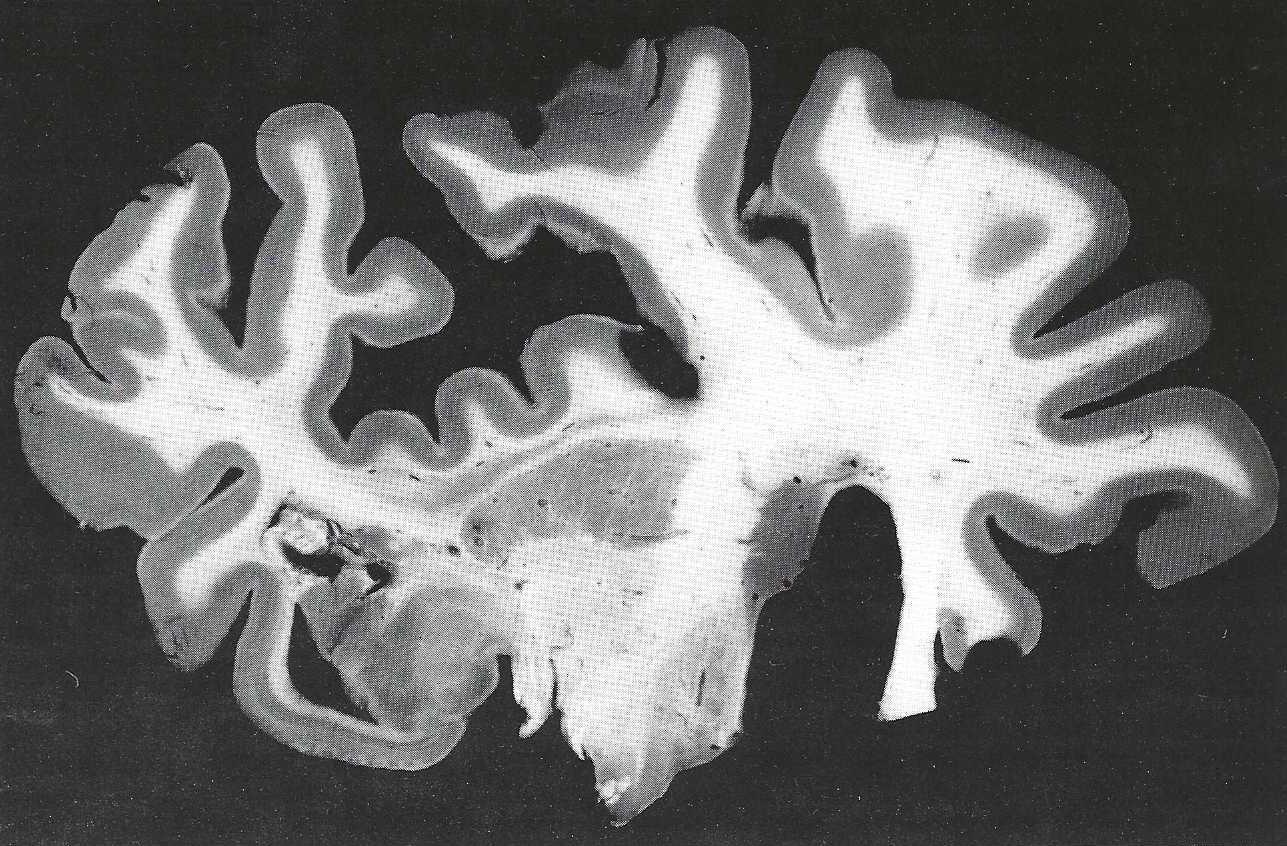 Figure 1. Coronal frozen section (2 mm) stained with astra blue and subsequently plastinated. |
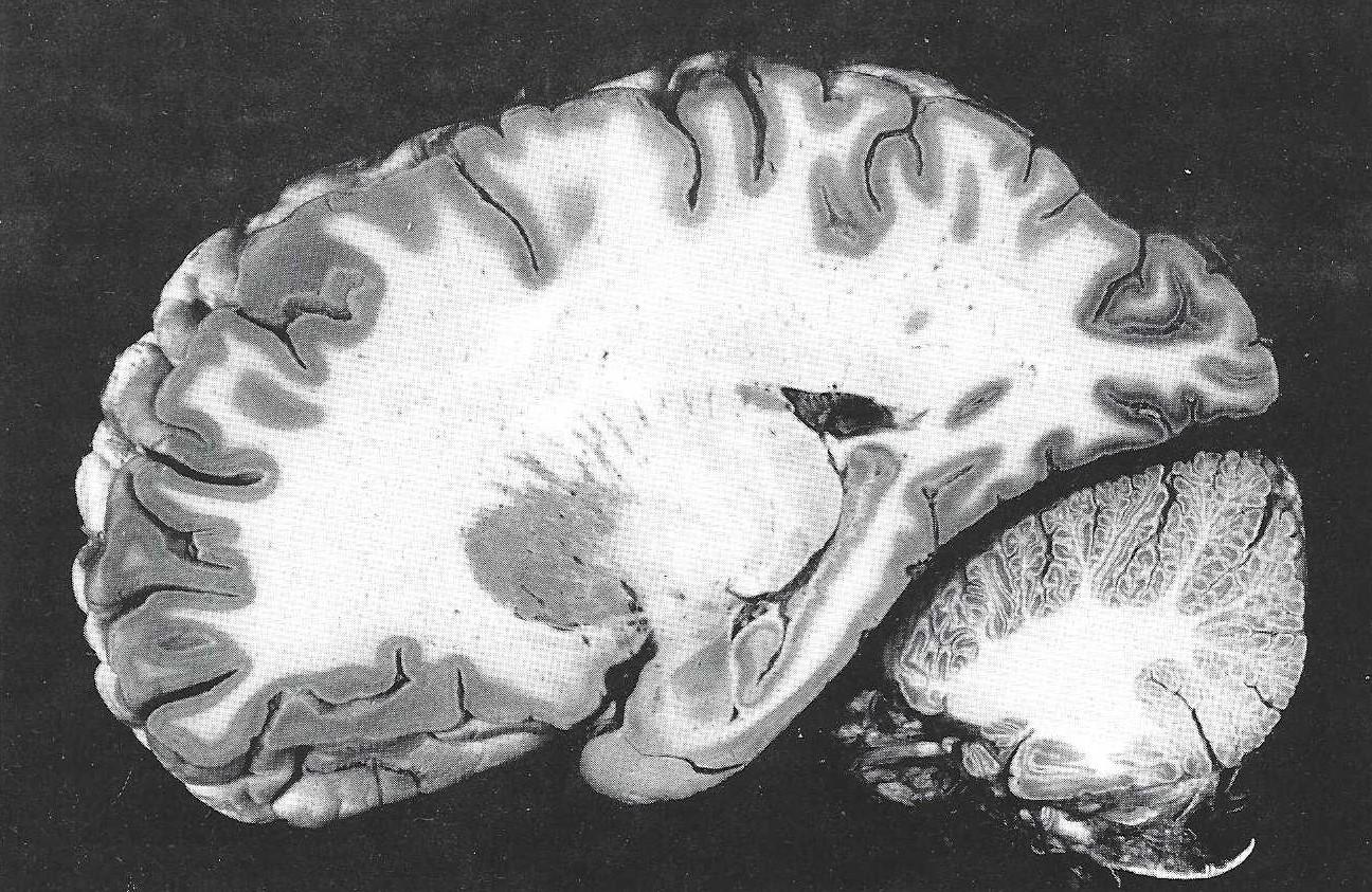 Figure 2. Sagittal macrotome section (1 cm) stained with astra blue and subsequently plastinated. |
Aldehydefuchsin demonstrates the distribution of lipofuscin granules in nerve cells of the adult brain. Varied lipofuscin content is visualized by different staining intensities (Figs. 3, 4). In this manner, a topographical delineation and the internal organization of nuclei can be studied by this pigmento-architectonic approach. (Braak, 1978a; 1984). The preparation of the aldehydefuchsin staining solution is complicated. Therefore, this staining procedure is much more complex than with astra blue. Commercially prepared aldehydefuchsin is not recommended since it may stain the white matter. Furthermore, it tends to produce artificial spots of intense staining in thick sections. For these same reasons, crotonaldehyde is used instead of paraldehyde, and pararosanilin is employed instead of fuchsin (Braak, 1978a). To obtain satisfactory results, only adult brains of more than 40 years of age should be used for the staining with aldehydefuchsin.
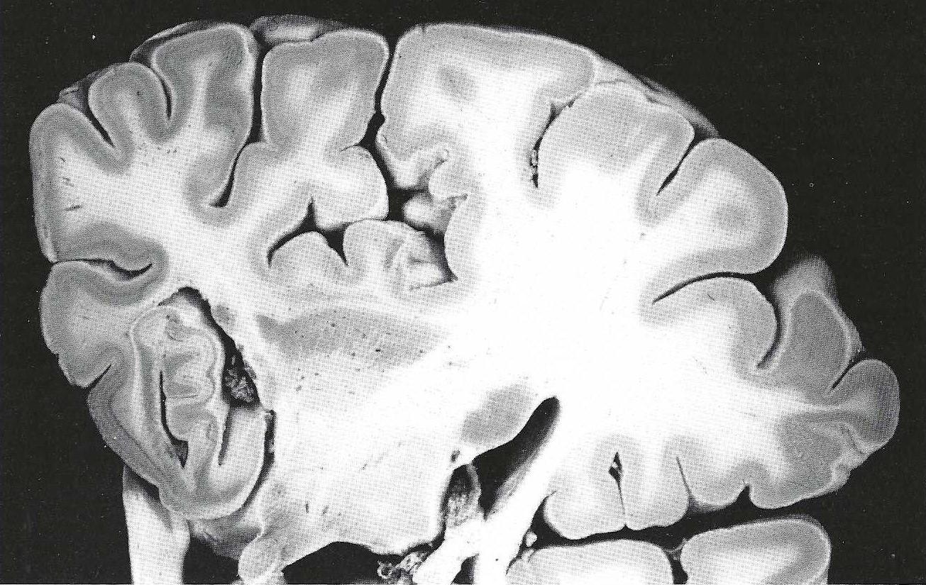 Figure 3. Coronal macrotome section (1 cm) stained with aldehydefuchsin and subsequently plastinated. |
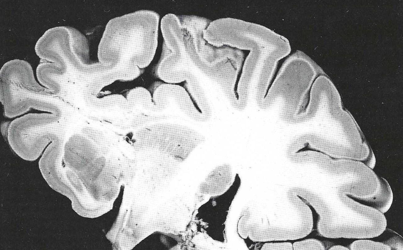 Figure 4. Coronal macrotome section (1 cm) stained with aldehydefuchsin and subsequently plastinated. |
FETAL HUMAN BRAIN TISSUE
Embedding fetal brain tissue in celloidin provides 0.2 - 2 mm thick sections. Other sectioning techniques (gelatin-embedded, polyethylene-glycol-embedded, or frozen tissues) are not applicable for fetal brain tissue because these sections do no withstand the staining procedure. Celloidin-embedding results in sections which shrink minimally and provide excellent preservation of vulnerable structures, such as in the septal region. These quality sections seemingly justify the relatively high expenditure of time which is required for the celloidin embedding procedure.
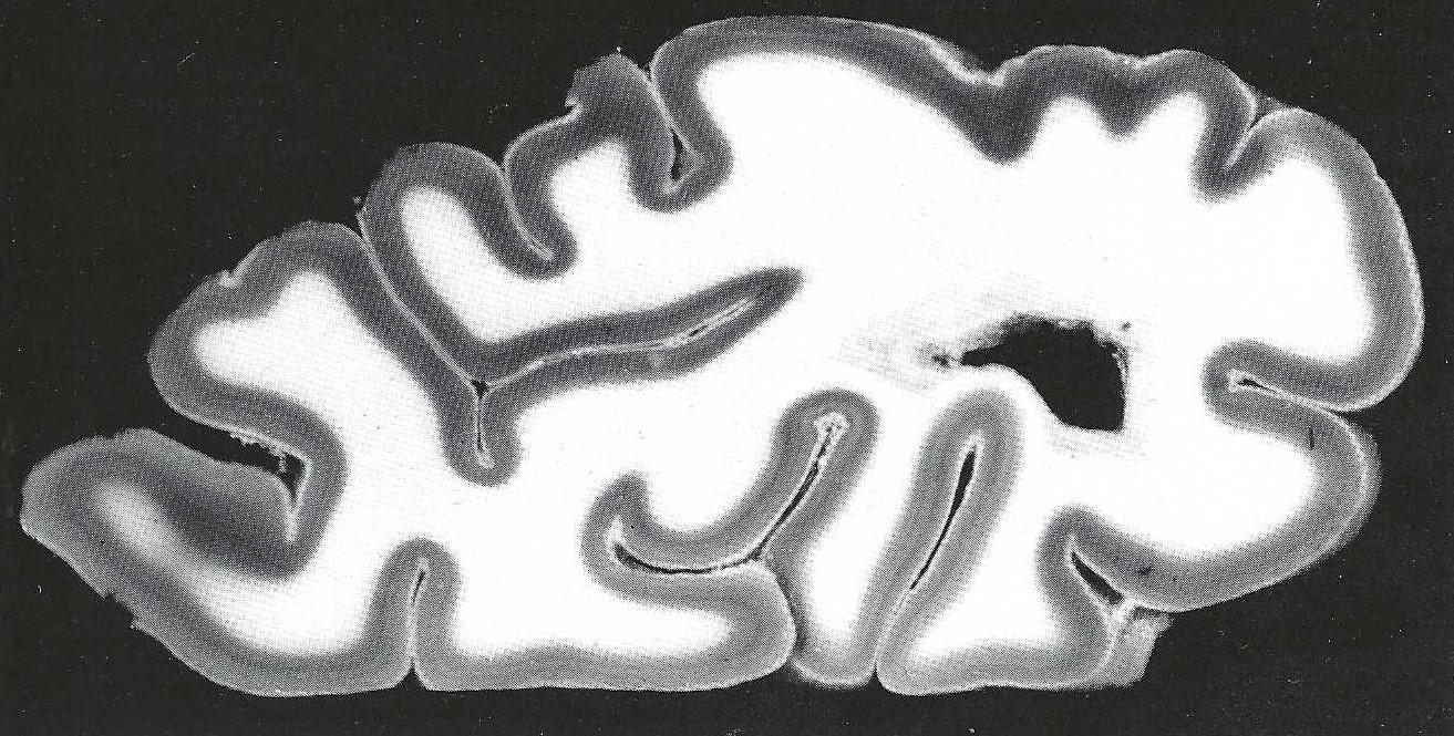 Figure 5. Fetal coronal section (embedded in celloidin,2 mm) stained with Darrow red and subsequently plastinated. |
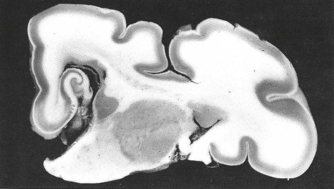 Figure 6. Fetal coronal section (embedded in celloidin, 2 mm) stained with Darrow red and subsequently plastinated. |
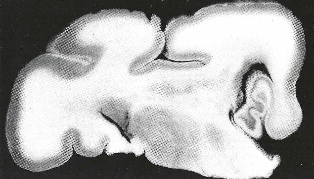 Figure 7. Fetalcoronal section (embedded in celloidin, 2 mm) stained with Darrow red and subsequently plastinated. |
|
The varied quantity of Nissl-substance within the nuclear greys are highlighted by Darrow-red, which is, to our experience, not possible by means of other Nissl stains in sections of these thickness. Darrow-red can easily be removed from the background of celloidin- embedded material (Powers and Clark, 1960; 1963). A gapless series of fetal brains can be cut frontally, horizontally or sagittally and stained. Hence, the architectonic organization of fetal brains at various developmental stages can be followed throughout developmental changes of the nuclear configurations (Ulfig, in press).
The standard S10/S3 plastination procedure is used for impregnation of fetal, as well as, adult brain sections (vonHagens, 1985). Dehydration of celloidin-embedded fetal brain tissue in 100% acetone removes the celloidin from the sections. Complete impregnation leads to flexible and opaque preparations with high tensile strength. Without much expenditure or experience this method can be established in any laboratory. Moreover, it should be stressed that material which has been stored for several years and which has lost its natural contrast between white and grey matter can be used.
ACKNOWLEDGEMENTS:
The author wishes to thank C.H. Medina for his excellent help.
Braak H: Eine ausfiihrliche Beschreibung pigmentarchitektonischer Arbeitsverfahren (A detailed description of pigmentarchitectonic methods). Mikroskopie (Vienna) 34:215-221,1978a.
Braak H: Simple and durable staining of thick sections of the human brain for macroscopic study. Stain Technol 53:87-89, 1978b. https://doi.org/10.3109/10520297809111447
Braak H: Architectonics as seen by lipofuscin stains. In: A Peters, EG Jones (eds.) Cerebral Cortex. Vol.1. Plenum Press, New York, pp. 59-104,1984.
von Hagens G: Heidelberg Plastination Folder: Collection of all technical leaflets for plastination. Anatomisches Institut 1,Universitat Heidelberg, 1985.
Mulligan JH: A method of staining the brain for macroscopic study. J Anat 65:468-472 , 1931.
Powers MM, G Clark: A note on Darrow Red. Stain Technol 38:289-290, 1963.
Powers MM, G Clark, MA Darrow, VM Emmel: Darrow-red, a new basic dye. Stain Technol 35:19-21, 1960.
https://doi.org/10.3109/10520296009114710
Romeis B: Mikroskopische Technik (P Bock ed.) Urban and Schwarzenbeck, Munchen Wien Baltimore, 1989.
Ulfig N: Technique for investigation the architectonics in human fetal brain tissue. Z mikrosk anat Forsch (in press).
Ulfig N, M Wuttke: Plastination of stained sections of the human brain. Anat Anz 170:309-312, 1990.