Department of Human Biology, School of Biomedical Sciences, Curtin University of Technology, Perth, Western Australia
Students enrolled in their third year of a Bachelor of Science course at Curtin University used plastination techniques to preserve their dissected specimens as part of the practical component of the embryology module of the Human Structure and Development unit. Students attended an information session on the methodology of plastination. To assess age, fetal pig specimens were measured and weighed. Students then chose developmental features they wished to portray.
Specimens were dissected by the students, dehydrated and plastinated. Progress was monitored by the students with respect to dehydration, shrinkage and color retention. After 8 weeks the plastinated specimens were examined and their component parts identified. Specimens were photographed with a digital camera and the resulting images imported into a HyperCard stack representing the work of the class. Images were labeled and linked to information about their age and sectioning. The plastinated specimens were retained for future student use.
Participation in the plastination gave the students responsibility for producing their specimens. Students learned specimen preparation combined with image digitization and multimedia presentation of material. Student evaluation of the plastination component of this unit and examples of their work are presented.
Presented in part at the 8th International Conference on Plastination, Brisbane, Australia, July 14-19, 1996.
Plastination, Embryology, Multimedia, Piglet
Gary Whittaker, Department of Human Biology, School of Biomedical Sciences, Curtin Uni- versity of Technology, P.O. Box U 1987, Perth 6845, Western Australia. Telephone: 61 8 9266 7995 / Fax: 61 8 9266 2339. Email: iwhittak@info.curtin.edu.au

![]()


The School of Biomedical Sciences offers students a range of degrees, including the BSc in Human Biology. These students choose their majors from three discipline streams; morphology, physiology and molecular genetics. Students in the morphology stream may select four modules to build their final third-year unit. One of these modules is called Human Structure and Development 331 and explores the structural changes which occur during embryological development.
Foetal pig specimens provide an inexpensive and easily-accessible animal model to study developmental changes and correlate with human development. Previously foetal pig specimens were used but the students had difficulty in relating their dissections to the lecture material in a once-off wet laboratory class. Therefore it was decided for the 1995 session to use plastination for preservation and to enhance the learning experience.
Educational rationale
Learning is a collection of activities whereby knowledge is constructed in the mind of the learners from resources presented to them. It is documented that students learn more effectively if engaged in some activity which provides an opportunity to practice the theoretical material from lectures and textbooks (Beard and Senior, 1980; Entwistle, 1981; Laurillard, 1981). Practical classes allow the students to manipulate material in three dimensions and to consolidate ideas which have been generated from reading or listening.
The use of fresh material is made less useful because it has a limited shelf-life. Preserving with formalin solution has the usual limitations associated with noxious fumes, and requires specific dissection-room facilities. The plastination process allows students to dissect the fresh specimens to show features which they wish to study then preserve them for later use. This reinforces their own understanding and illustrates concepts when working with other students in a collaborative learning tutorial. The plastinated specimens can be used in ordinary classrooms to expand the learning experience of practical work beyond the practical sessions.
In our example, students are actively involved in the selection, preparation and presentation of the specimens rather than merely passively viewing specimens prepared by someone else. Students select their own area of interest to study and are motivated by identifying various components displayed, and understand their functional relationship with surrounding structures.
Students attended an information session on the methodology of plastination which was designed to relate to their experience and knowledge of histological techniques. Foetal pig specimens of various stages of development were obtained from a local abattoir. Students examined the specimens, measured and weighed them to determine their various ages. Subsequently they decided which aspects of development they wished to portray in their specimen. Stu- dents consulted with the lecturer and the technical expert to reach a decision. Some students opted for equivalent sections of specimens at different stages of development while others chose to section and prosect one specimen to trace detailed structures at a particular age.
Dissection and sectioning was carried out on the fresh specimens while some were immediately frozen to facilitate sectioning. Smaller specimens were sectioned at room temperature using a brain knife and the frozen specimens were sectioned with a band saw. Sectioning was carried out or supervised by technical staff but at all other times students were directly involved in the preparation of their specimens. Specimens were then plastinated using the S10 technique (von Hagens, 1985).
Fixation and dehydration
To accelerate the process the specimens were freeze- fixed (von Hagens, 1985). This ensured that the specimens would be completed within one semester. As an additional advantage, freeze fixation preserves colour better than the traditional fixation and dehydration method. Specimens were submerged in the first acetone bath containing 5% formalin (stabilized with 10% methanol) for two weeks. They were then dehydrated by freeze-substitution in 100% acetone. Both fixation and dehydration were preformed at -25°C. Dehydration continued for 4 weeks until the specimens had a water content of less than 1%. Progress was monitored by the students on a weekly basis with respect to dehydration, shrink- age and colour retention.
Forced Impregnation
Specimens were immersed in polymer (Biodur S10+S3) which was placed inside a vacuum tank in a freezer (-25°C). The specimens were left overnight to equilibrate with the polymer. A vacuum was applied to the impregnation bath and pressure was gradually decreased to 10mm Hg over a three weeks period.
Curing
After impregnation the specimens were drained of excess polymer and then cured with Biodur S6. Due to time constraint, the fast-cure method was adopted as the preferred curing method. The specimens were placed in a sealed chamber with the volatile curing solution (S6). After three days the specimens were dry to touch, allowing the students to handle their specimens quickly after impregnation. The specimens were then stored in a sealed container along with desiccant for three months to allow complete curing.
After 8 weeks from the commencement of the project the plastinated specimens were examined and their component parts identified. The specimens were photographed with a digital camera and the resulting images imported into a HyperCard stack called Piglet 95, which represented the work of the class. Students labeled the images and linked this to information about the age of the piglet specimen and to the plane of sectioning.
After the plastination process was completed, the technical success of the specimens was evaluated. A total of 31 pieces were deemed useful for the project (figure 1), representing 82% of the total prepared prosected material. Specimens were considered a failure if they were either incompletely impregnated or fragmented from their original specimen.
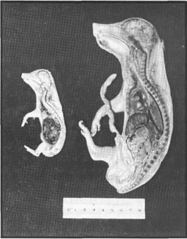
Figure 1. Plastinated 20 (left) and 60 (right) day foetal pig specimens. Midsagittal sections
Good colour differentiation was exhibited by all specimens. Thoracic and abdominal viscera in particular showed extremely good colour differentiation. In some cases the adipose tissue within the abdominal cavity even retained it's yellow colour. Nervous tissue however was less successful. Shrinkage was quite marked and in many cases the brain, especially in the smaller specimens lacked the detail expected.
Evaluation of outcomes
The plastination process and subsequent HyperCard stack contributed to 20% of the semester mark. Each student was assessed with regard to their planning and execution of specimen preparation rather than the success of the subsequent plastination. This was the first time plastination of piglets had been attempted in this unit, therefore little was known of the likely success of such preparations. Much of the technical work was supervised or carried out by staff, and there were no guidelines given to students as to the most suitable specimens to plastinate.
In informal focus groups during and at the end of the semester, students spoke of the plastination procedures with enthusiasm. While some of this could have been due to the novelty of doing something which had not been tried before, the combination of practical experience and theory was supported with the idea that the students would have "some- thing to take away with them" at the end of the semester. Negative comments related to the time lag between the prosection of the piglets and the final curing of the plastinated specimens and the time constraints of the S10 procedure.
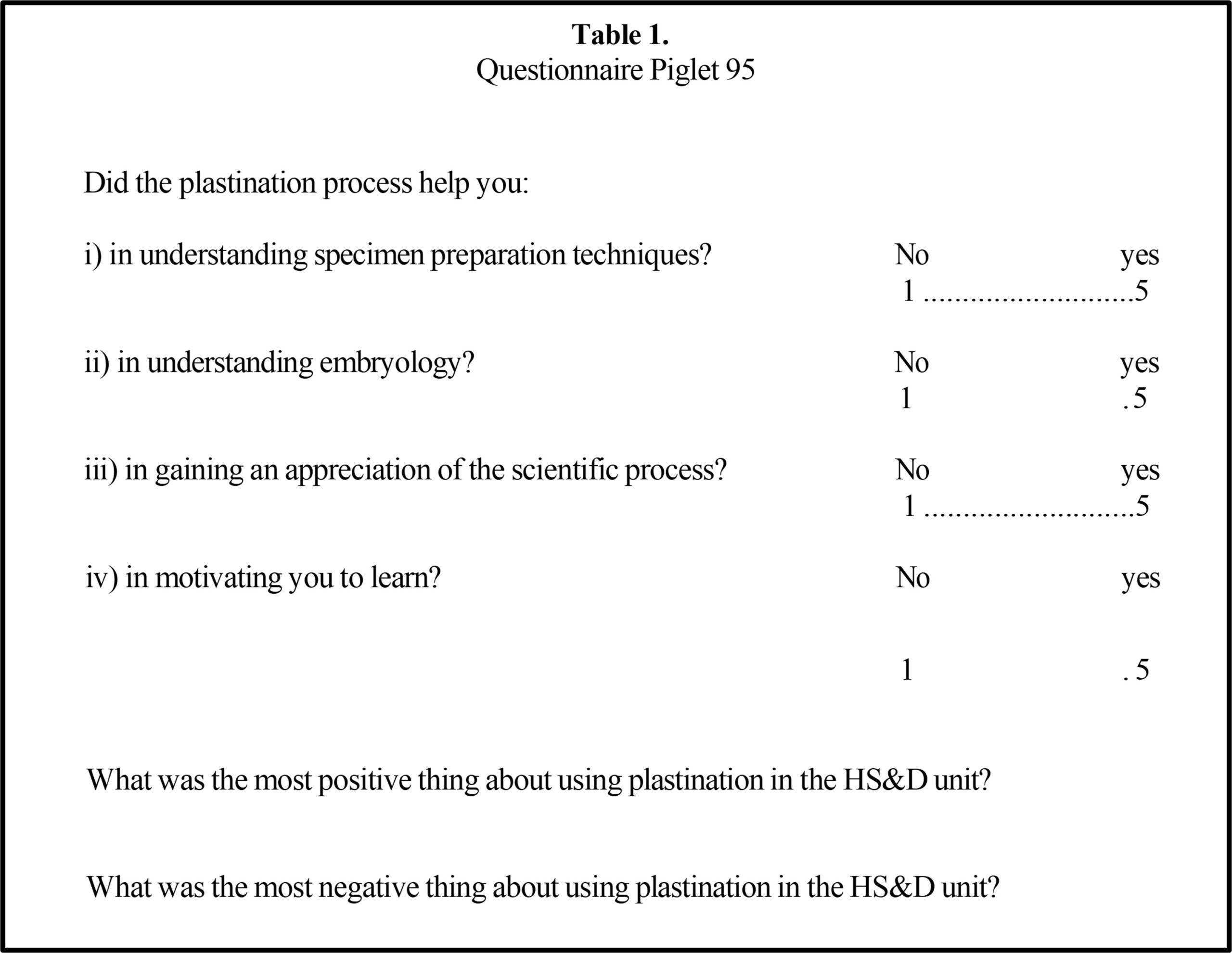
Table 1.
Questionnaire Piglet 95
A sample of students (n=4) which represented 40% of the class were interviewed after the semester was over, and asked to reply to a questionnaire (Table 1). Unfortunately the class, having completed their degrees, had dispersed after their final examination and our sample was restricted. Students replied favorably to the use of plastination within the unit. They found plastination "useful in helping them relate embryological structures in three dimensions and relating them to the changes which occur during foetal development". The students also wanted to be "more involved in the actual technical process giving them more responsibility of the actual plastination of their own specimen".
Peer review assessment was conducted with the assistance of staff in the School of Biomedical Sciences and in the Veterinary School at Massey University in New Zealand, where plastinated sheep brains are used in teaching. Infor- mal focus group sessions were used to share experiences and exchange ideas, but no report on this process has yet been published.
Future Directions
Due to the success of this trial, plastination will be retained in the unit Human Structure and Development 331 at Curtin University. However, as a result of the evaluations conducted and of the trial of alternative plastination techniques, some changes will be made to the timing and certain dissections will be suggested to ensure greater success of the plastination portion of the project.
The S10 process in preserving student specimens certainly proved to be useful and successful. However, due to the length of the process and the time constraint within which we worked, it was felt that if this were to be attempted again the students would have to engage in their selection and dis- section of specimens much earlier in the course or alternatively a shorter plastination process be used.
An alternative process, the E12 technique (von Hagens, 1985; Weber and Henry, 1993), has been tried, and has been very successful in showing great detail of foetal structures. The use of this method will have many advantages over the S10. Although technically more challenging than S10, it could allow a shorter processing time. This reduces the time lag between sectioning and end product, and thus will allow students to work more extensively with their plastinated specimen. Of all the prosections tested, more students opted for sectioning their specimens, and these sections proved to be very successful, E12 works extremely well for sectioned specimens. E12 plastination sections are thinner which al- lows greater topographical investigation of developing viscera within the foetal pig. Hollow organs are preserved in E12 but are often lost using S10.
With more time for development of ideas using the specimens in class, plastination in the future will further enhance the learning experience. It will also allow for increased self- directed learning as these specimens can easily be used in the library or at home. In the future, students will be more directly involved during the technical procedures of plastinating their specimens. This will increase their perceptions as stakeholders in their learning.
Plastination of foetal piglet specimens allows students to relate to histological and basic morphological techniques: prosection, resin embedding and progressive dehydration. It reinforces concepts of the scientific method and enhances the learning of a difficult section in morphology - embryological development. The generation of a HyperCard stack of images, to illustrate the students' input into a tangible representation of their learning, was an important adjunct to the plastination process.
The HyperCard stack, Piglet 95, can be viewed at the Human Biology Multimedia website:
http:// www.curtin.edu.au/curtin/dept/biomed/teach/hubiol/hb.html
Beard RM, Senior IJ: Motivating Students, Routledge and Kegan Paul, London, 1980.
Entwistle N: Styles of Learning and Teaching, John Wiley, New York, 1981.
Harel I and Papert S (eds): Constructionism, Ablex Publish- ers, Norwood, New Jersey, 1991.
Laurillard DM: The Promotion of Learning Using CAL, Computer Simulation in University Teaching, (ed. Wildenberg D), North-Holland Publishing Co, Amster- dam, pp 83-90, 1981.
von Hagens G: Heidelberg Plastination Folder: Collection of all technical leaflets for plastination. Anatomische Institut 1, Universitat Heidelberg, Heidelberg, Germany, 1985.
Weber W, Henry RW: Sheet plastination of body slices - E12 technique, filling method. J Int Soc Plastination, 7 (1): 16-22, 1993. https://doi.org/10.56507/EZGX2343