1- University of Virginia Medical Center Department of Pathology Charlottesville, Virginia
2- Mercer University School of Medicine Department of Pathology Macon, Georgia
DNA ploidy analysis by flow cytometry is gaining widespread use in the study of neoplastic disease. Although we have observed satisfactory preservation of light microscopic detail in plastinated tissue, we questioned whether plastinated neoplasms were still suitable for ploidy analysis.
DNA; Plastination; Silicone; S10
H Frierson University of Virginia Medical Center Department of Pathology Charlottesville, Virginia

![]()


DNA ploidy analysis by flow cytometry is gaining widespread use in the study of neoplastic disease. Although we have observed satisfactory preservation of light microscopic detail in plastinated tissue, we questioned whether plastinated neoplasms were still suitable for ploidy analysis.
A representative sample was taken from silicone rubber-impregnated squamous carcinoma of the esophagus and a silicone rubber-impregnated adenocarcinoma of the colon. The samples were immersed in 5% sodium methoxide dissolved in methanol for 48 hours to remove the silicone polymer. They were then washed in fresh methanol and divided in half. One half of each sample was submitted for DNA ploidy analysis without further processing. The other respective halves were placed in cassettes and subjected to routine tissue processing on an automated tissue processor, whereafter they were embedded in paraffin. A 50 micron section from each of the two paraffin blocks was placed into separate glass centrifuge tubes and deparaffinized according to the technique of Hedley et al (1). Briefly, sections were dewaxed with xylene and rehydrated with decreasing concentrations of ethanol (100, 95, 70, 50%). After washing the cells in distilled water, 1 ml of 0.5% pepsin (Sigma P-7012, St. Louis, MO) in 0.9% NaCl adjusted to pH 1.5 was added. Tubes were incubated in a waterbath at 37° for 45 minutes and vortexed intermittently. IxlO6 cells were then placed into a DMSO-citrate buffer, frozen rapidly, and stored at -80°C.
The deparaffinized cells were prepared for cytometric analysis using the detergent- trypsin method of Vindelov (2). After staining with propidium iodide, nuclei were passed through a 70 um nylon filter and analyzed with an Epics C flow cytometer (Coulter Electronics, Inc., Hialeah, FL) equipped with a 5-watt argon laser. Excitation of propidium iodide occurred at 488 nm and the fluorescent emission was measured above 590 nm. Twenty thousand nuclei from each specimen were analyzed.
Non-neoplastic nuclei present in the paraffin blocks served as an internal diploid standard. Tumors were considered to be DNA aneuploid when another GO-G1 peak was present in addition to the diploid peak.
The DNA index was calculated as the ratio of the mean channel number of the GO-G1 peak for the neoplastic nuclei to the mean channel number of the GO-G1 peak for the diploid nuclei. The coefficient of variation (CV) was calculated for each GO-G1 peak using the half maximum-peak height.
Both deplastinated, non-processed sections were unsuitable for flow cytometric analysis as the constituent cells resisted the disaggregating procedures. The deplastinated, processed and paraffin embedded material gave much more satisfactory results.
 Figure 1 |
 Figure 2 |
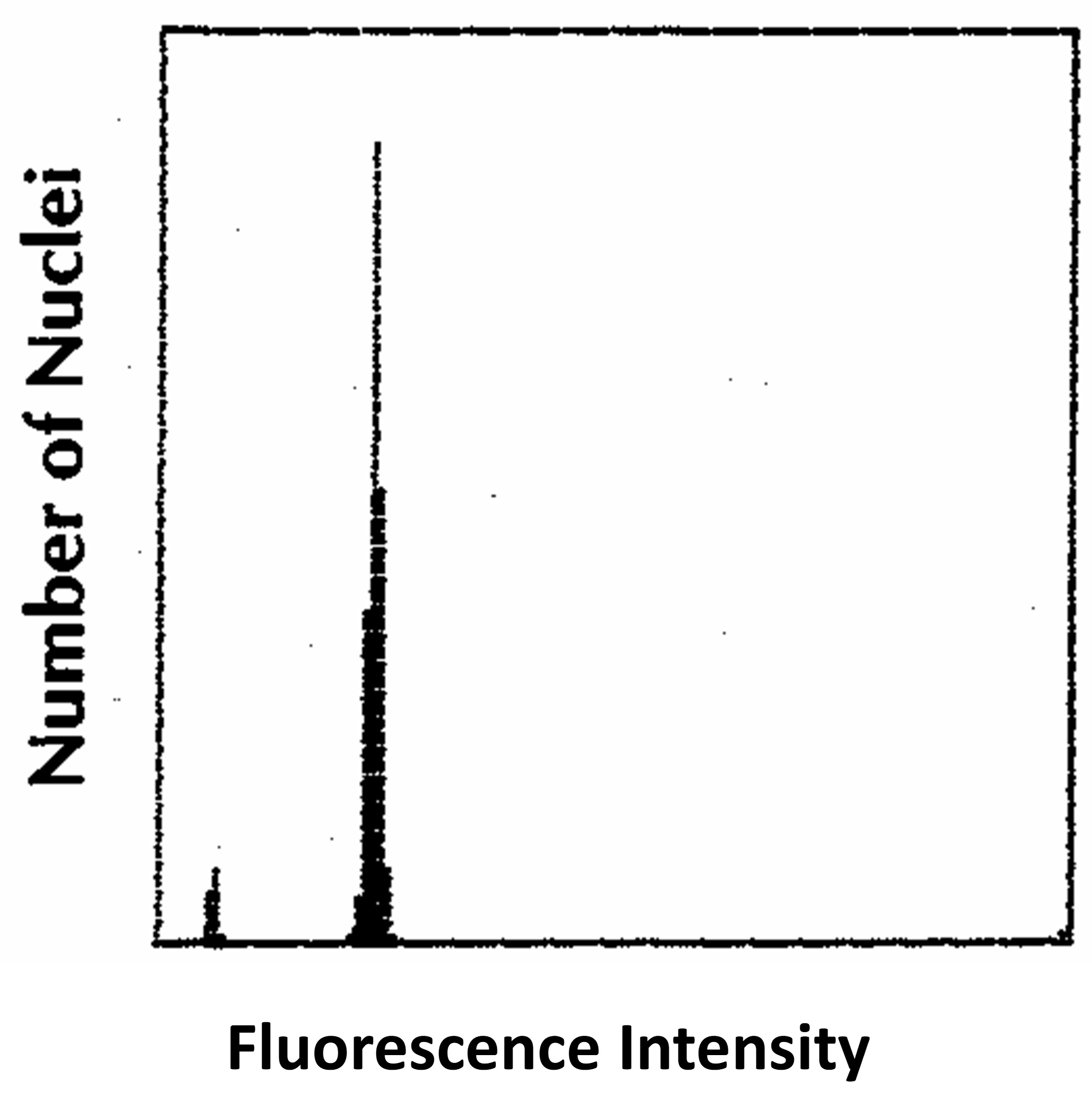 Figure 3 |
Figure 1 is a histogram of the nuclei from the esophageal squamous carcinoma, showing only a diploid peak. Figure 2 is a histogram of the nuclei from the colon i c adenocarcinoma, showing both aneuploid and diploid peaks; the DNA index is 1.27. Both histograms contain abundant debris concentrated in the hypodiploid region. The coefficient of variation for each of the DNA diploid peak is less than 10%.
Figure 3 is a control histogram of the nuclei from fresh peripheral blood lymphocytes showing a single narrow diploid peak.
From this preliminary study we concluded that it is possible to perform subsequent ploidy analysis on plastinated tissues. Plastinated tissue does not appear to be optimal material for ploidy analysis, but can be used successfully if no other portion of a specimen is available.
1- Hedley DW, Friedlander ML, Taylor IW, Rugg CA, Musgrove Method for analysis of cellular DNA content of paraffin-embedded pathological material using flow cytometry. J Histochem Cytochem 31:1333-1335, 1983.
https://doi.org/10.1177/31.11.6619538
2- Vindelov LL, Christensen IJ, Nissen A detergent-trypsin method for the preparation of nuclei for flow cytometric DNA analysis. Cytometry 3:323-327, 1983. https://doi.org/10.1002/cyto.990030503