Orthopaedic University Hospital, University of Heidelberg, Germany
In the investigation of osteoarthritis and degenerative tendinous changes, the sheet plastination method (E 20) is used routinely. The described method allows the demonstration of microvascular anatomy and pathology following arterial perfusion with epoxy resin. As compared to other investigations on human cadavers, the plastination method is capable of demonstrating subchondral yascular changes and their relationship to cartilage defects and early subchondral sclerosis.
Plastination, Achilles tendon, Osteoarthritis, New anatomical preparation techniques.
Graf, B Orthopaedic University Hospital, University of Heidelberg, Germany

![]()


The method of plastination, inaugurated by von Hagens in Heidelberg, has been used routinely for the last five years in the investigation of the role of the paratenon in the blood supply of tendons (Graf et al., 1990) and in the etiology of early osteoarthritic changes of the patella (Graf et al., 1988; Schneider et al., 1989). The advantages of this method are the increased durability of the prepared specimens, ease of morphometric examination and minimal damage to the plastinated tissue.
Between 1985 and 1990, 16 human knee joints and 12 Achilles tendons were examined using the plastination method as described by von Hagens (1979). After dissection of the femoral artery and vein, a catheter was inserted into the artery and the plastination compounds BIODUR E 20/E 2, after mixing, were infused into the vessel using a continuous pressure of 130 mm Hg. After freezing, at -70 "C, the specimens were cut into 2 mm thick slices on a band saw and plastinated in sheets using the draining technique as described by von Hagens (1987). The plastinated sections were placed between polyester foils and glass plates and allowed to cure. Later, they were examined macroscopically, as well as, with the light microscope.
Using this method, extensive lesions were demonstrated at the osseochondral border and within the cartilage itself, while the surface of the cartilage appeared undisturbed and smooth. There was a marked increase of subchondral neovascularization and sclerosis (Fig. 1).
Regarding the vasculature of the Achilles tendon, a large number of anastomoses were noted between the extratendineous and intratendineous vessel systems. Within the extratendineous system, there was a remarkable number of small blood vessels as compared to the tendon itself. Four to five centimeters proximal to its insertion at the calcaneus, there was a zone of relative hypovascularization within the Achilles tendon. The paratenon in this area was unremarkable. There were no connecting blood vessels between the calcaneus and the distal parts of the Achilles tendon (Fig. 2).
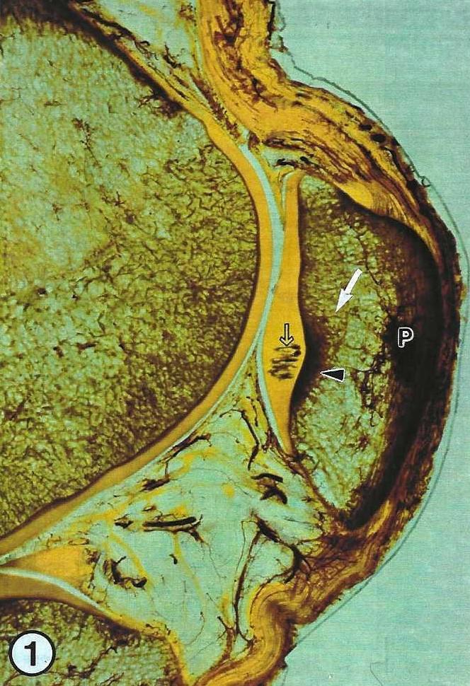 Figure 1. Human patella (P), cut transversely. Note the retropatellar degenerative changes (open arrow) on the caudal surface of the patella or cartilage, and the marked sclerosis (arrowhead) and the hypervascularization (arrow) of the subchondral area. |
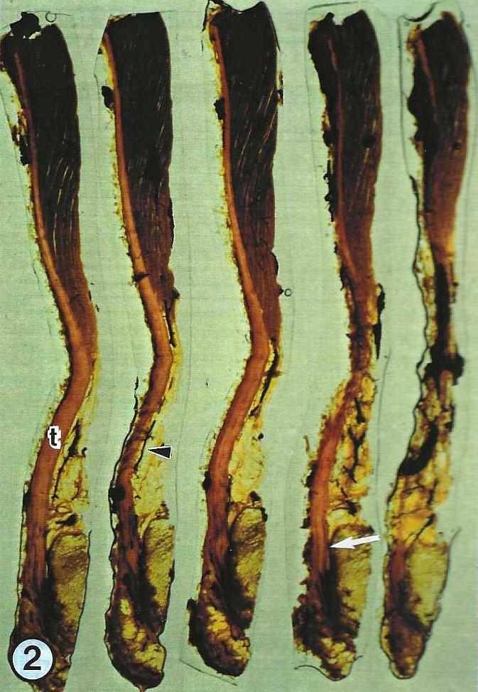 Figure 2. Longitudinal sections of a human Achilles tendon (t). Note the well vascularized paratenon (arrow head) as compared to the tendon itself. No connective vessels are seen to cross the osseochondral insertion (arrow). |
For the first time, this method has been applied in the field of osteoarthritis research. It has been possible to show the relationship between degenerative changes within the cartilage, subchondral vascularization and subchondral sclerosis. These results hint at possible roles of the intraosseous area and, especially, the subchondral area in the early developmental stages of osteoarthritis (Graf et al., 1989a). These morphological findings correspond to clinical stage I chondromalacia.
The most common site of rupture of the Achilles tendon lies 4-5 cm proximal to its insertion, the site of its weakest vascularization. This, however, does not prove conclusively a relationship between poor vascularization and rupture site, as there are no ruptures of the tendon at its insertion to the bone, where no blood vessels are found (Grafetal., 1989b).
Graf J, U Schneider, FU Niethard: Vascularizacion subcondral y osteoarthritis. XXV Congreso Argentine de Ortopedia y Traumatologia, Buenos Aires, 1988.
Graf J, E Neusel, FU Niethard: Die Bedeutung der subchondralen Vaskularisation fur die Entstehung posttraumatischer Chondromalazien an der Patella. Aktuelle Unfallheilkunde 5/6, Schnetztor-Verlag, p. 340-342,1989a.
Graf J, U Schneider, FU Niethard, G yon Hagens: The microcirculation of the Achilles tendon - demonstrated by the plastination. XIII Federative International Congress of Anatomy, Rio de Janeiro, 1989b.
Graf J, U Schneider, FU Niethard: Die Mikrozirkulation der Achillessehne und die Bedeutung des Paratenon. Handchirurgie 3:163-166, 1990.
Schneider U, J Graf, A. Gttfbacher, FU Niethard: Comparative morphological and arthroscopic investigations in chondromalacia of the patella. Sandorama IV:24-26, 1989.
von Hagens G: Impregnation of soft biological specimens with thermosetting resins and elastomers. Anat Rec 194:247- 256,1979.
https://doi.org/10.1002/ar.1091940206
von Hagens G: The current potential of plastination. Anat Embryol:411-421,1987.
https://doi.org/10.1007/BF00309677