1Conservateur du Musée Fragonard École Nationale Vétérinaire d’Alfort, Maisons-Alfort, France
2 Division of Biomedical Sciences (Anatomy), St. George’s, University of London, London, UK
Dr. Louis Auzoux (1797-1880) is well known for the anatomical models of papier mâché that he produced and exported all over the world. Although the human models are more widely known, they are by no means the only ones that the famous medical industrialist designed and marketed: animals, plants and especially flowers are another facet of his art. Models of the horse were especially important for Auzoux’s business. The paper horses, the sets of bone defects and jaws that he created were purchased in great quantities by the French government of the day to provide the materials needed for training recruits in a time of war. There was also a programme to improve horse breeding throughout France through these fascinating objects. These magnificent creations that were distributed all round the world, and which once were the pride of France, are now damaged, ignored and dispersed. Sadly, they are now in great danger of being lost forever. This historical review is an extensively revised translation of an article that was originally published in French (Degueurce, 2013).
Auzoux, anatomical model, papier mâché, horse, honeybee, silkworm
PJ Adds, Institute of Medical and Biomedical Education (Anatomy), St George’s University of London, Cranmer Terrace, London SW17 0RE UK. Tel: +44 (0) 208 725 5208, email padds@sgul.ac.uk

![]()


The mannequins of Dr Auzoux, an industrial success in the service of veterinary medicine
The discovery in the National Archives of France of a hitherto unexploited archive (Arch. Nat.) followed by several visits to Saint-Aubin-d’Ecrosville, where Auzoux’s factory was established, and conversations with the curator of the Neubourg Museum 1, have shed new light on the life and legacy of this most industrious, industrial doctor. This article summarises the authors’ research into Auzoux’s achievements with the domestic animals (Degueurce, 2013).
Preservation and decay, the bane of the anatomist’s life
Ever since the first anatomists attempted to explore the body to reveal its structure, they despaired as their careful dissections withered and, inevitably, decayed.
Transforming the ephemeral into the durable became an urgent priority, particularly in the second half of the 18th century, when the demand for anatomical education became much greater.
Many museum collections from this period display the attempts that were made, which mainly fall into two categories. The most common consisted of preserving the whole body, or its parts, by dehydration or immersion. Dehydration, or mummification, had the advantage of eliminating cellular fluids, and thereby preventing putrefaction. While Honoré Fragonard2 remains the best known practitioner of this technique, there were many others who also used it to enrich the museum collections of Faculties of Medicine and ‘cabinets of curiosities’ throughout Europe. This method, however, had the drawback of reducing even the most bulky muscles to thin, desiccated strips. Smaller specimens could be submerged in a preserving bath, which preserved their bulk, but it was difficult to use this technique for whole-body specimens.
The second approach was to make a model – an artificial representation – of the specimen before it decayed, and this provided a challenge to the modellers, artists and craftsmen of the day. Many different materials were tried, including coloured wax, plaster and even glass (Degueurce 2012a, p. 80). But the material that achieved a global success in the 19th century – then only to fall into total oblivion – was papier mâché. And papier mâché will always be associated with the name of Louis Auzoux (1797-1880), who exploited its possibilities so well that he created a flourishing industrial enterprise, producing anatomical models that, to this day, can still be seen in the museums of five continents.
Louis Auzoux and his papier mâché anatomical models
Louis Auzoux, doctor of medicine and brilliant inventor, was born in 1797 into an affluent family of cultivators in the village of Saint-Aubin-d’Ecrosville, about 100 km West of Paris (Degueurce 2102b, p. 23-34). His outstanding academic achievements, which led to a doctorate of medicine in Paris, gave him access to the medical celebrities of the day. The exact circumstances which pushed him into launching himself into “the anatomical industry” remain unclear, though what is certain was his talent for self-publicity.
We know that he started his researches very early on, and we know that his work cost him a great deal - financially as well as intellectually. Originally, he was inspired by Jean-François Ameline, Professor of Anatomy at Caen, who enjoyed a modest success with a novel type of anatomical mannequin, made from pieces of card fixed on to a real skeleton. These mannequins could be dismantled layer by layer, tracking the nerves and vessels and revealing the anatomical relations of the abdominal and thoracic viscera (Ameline, 1825 p. 5). This ingenious idea was, however, rather limited, and the small number of parts meant that it was a long way from being an adequate substitute for a real dissection.
Auzoux, then a young student in Paris, became aware of the process, and even travelled to Caen to visit Ameline’s workshop. A short while after, in September 1822, he presented to the Academie royale de médecine his own version, a “membre abdominal” with real bones for its base, just like Ameline’s, making them direct competitors (Collectif, 1825, 1). A new piece – a head, neck and superior part of the trunk – soon attracted the attention of the Government (Collectif, 1825, 2), which then placed an order for a whole body mannequin.
Delivered in 1825, this piece contained an innovation that would revolutionise its production: he used artificial bones instead of the real skeleton. So began for the doctor a fruitful career that led him to create several hundred models, produced quasi-industrially, at an affordable price which assured their wide distribution. This prototype was revised, and led to the grand modèle of 1830, which would be marketed, incredibly, right up to the 1970s. There followed an anatomised female in the position of the Venus of the Medicis, then numerous models of organs, singly or assembled to form a region of the body. In 1834, Auzoux dubbed his invention ‘anatomie clastique’, (clastic anatomy) from the Greek klaeïn, to break up/separate, an allusion to the educational dismantling of his various specimens (Anon. 1834, p. 453).
Nor did he stop at Man. As an eclectic naturalist and keen zoologist, Auzoux also produced models of ’type specimens’ of a variety of animals, including the turkey “as the type specimen of the fowls”, a shark “as the type specimen of the cartilaginous fishes”, and the cockchafer “as the type specimen of the adult insects”. The series was completed with the sea perch (fishes) the leech (annelid worms), and the boa constrictor (reptiles).
Particular species of animals that were important for the economy would become the object of even more detailed models. For the bee, Auzoux created a virtuoso production, showing, on a honeycomb, the stages of development and the internal structure of the adults. There followed the silkworm, with its butterflies of both sexes and their caterpillars. As for the horse, it was to hold a place of prime importance in the life and work of Louis Auzoux, just as it did in the lives of his contemporaries: draught animal, pivot of industry and agriculture, animal of luxury for the haut monde, and indispensable auxiliary of the army. It is easy to forget how central the horse was to life in the 19th century, and the interest that it sustained was universal, so it is not surprising that the idea of an equine model followed soon after his first human models.
The creation of the first equine model
The anatomical horse, which, as we shall see, was extremely complex, occupied Louis Auzoux almost from the beginning. Documents in the archive track the progress of the project from its beginnings in 1842 in the form of an exchange of letters with a young relative, an officer cadet at the Royal Cavalry School of Saumur3 in Western France. This young cadet informed Auzoux that his Professor of Hippology, the Marquis de Saint-Ange4, thought that a great advantage could be gained by using an equine mannequin based on the human papier mâché model that was already in use at the School. The anatomical specimens they were using, dehydrated and tarred5, were in poor condition, and could give only a basic idea of the anatomy of the horse. Two years later, Louis Auzoux wrote back, describing in detail the famous horse, apparently now finalised; in his letter he explains the considerable difficulties he had to overcome, in particular from the lack of accurate anatomical illustrations (Arch. Nat). He envisaged collaborating with the Professor to produce a simplified, less expensive version adapted for a wider distribution among soldiers in the ranks.
To make his horse model, Auzoux first had to carry out a completely original anatomical study, just as he had done earlier for the human model. The file on the equine model preserved in the National Archives confirms the paucity of information that was available at that time, and includes just one plate from the celebrated Cours d’hippiatrique by Philippe-Étienne Lafosse (Lafosse, 1772) (Fig. 1).
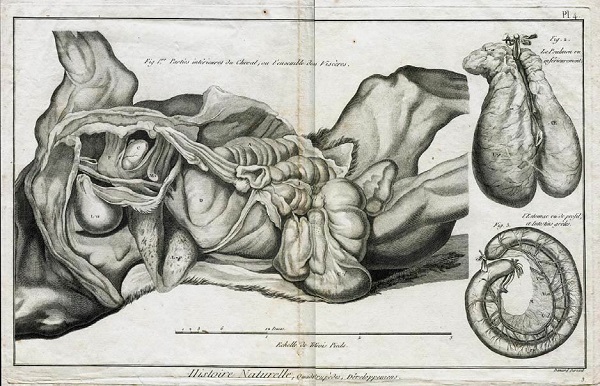
Figure 1 - Plate showing three engravings taken from the Cours d’Hippiatrique by Phillippe-Étienne Lafosse (1772).
Animal anatomy was still a developing science, and there was still much to learn about equine anatomy. Auzoux describes his model thus:
The horse is at rest. I have taken the pose and the balance from the training manual of M. Lecoq, professor at the veterinary school of Lyon. For the anatomical details, I have had to reproduce them after nature for there no longer exists a complete anatomy of the horse: the treatise of M. Rigot6 was helpful, but it covers only the bones and muscles”.
Auzoux had to wait two more years before he could submit his model to the Royal Academy of Medicine (Renault, 1845). He also sought the judgment of his fellow professionals: the archives contain several notes, jotted down while veterinary teachers examined his specimen, as well as numerous letters agreeing to meetings for these evaluations. Auzoux greatly valued their contribution:
“While occupied with the horse, I have had of necessity to make numerous loans to the veterinary schools, either for the classic design of the horse, or for the changes introduced in certain organs” (Auzoux, 1858, p. XIV).
In the field of agriculture and breeding, Auzoux benefitted greatly from the friendship of Antoine Richard (Richard du Cantal)7, an extraordinary character: a veterinarian but also doctor, farmer, agronomist, one time teacher and Director of the School of Stud farms, before following a political career. Richard published several important works on the improvement of the horse, in which he stressed the importance of the structure and mechanics of the animal’s body (Richard, 1847).
Auzoux was introduced to the military world by Colonel Maxime Jacquemin, Second in Command of the School of Cavalry8. As a young man during the last military campaigns of the Empire, Jacquemin had been struck by the lack of training the recruits received in caring for their horses, with deplorable consequences for these unfortunate animals. For him, hippology was an exact “mathematical” science, based on the structure and function of the body of the horse, hence his high opinion of Auzoux. Jacquemin carried on an extensive correspondence with the doctor-industrialist, on the subject of horse anatomy, the hoof, and the models showing limb defects (Arch. Nat.)
The different horse models
Auzoux’s first specimen9, presented to the Royal Academy of Medicine in April 1844, was described as “an equine of 1.1 metres in height (to the withers)”. Following the same principle as his human écorchés, the right part displayed superficial structures while the left half could be taken apart to reveal deeper structures. The trunk was split horizontally. Having first removed the limbs, the dorsal portion, complete with head, neck and viscera could be raised in one piece via a hinge placed under the tail (Figs. 2a-e) (Dumont et al., 2008).

Figure 2a - Right side of the horse, showing the vessels and the superficial muscles; one can clearly see the plane of cleavage between the dorsal and ventral parts allowing the horse to be opened.
The inspectors of the Academy of Medicine noted several imperfections: the mannequin was clumsy: “too wide in the chest”, the legs were “too bulky”; the muscles of the posterior regions were “too massive”. But all were in agreement in praising both the initiative and the result. Auzoux had succeeded.
In 1845, he marketed his cheval complet (whole horse), complete with a booklet listing all the removable parts, and describing how they could be removed, and a list of the countless anatomical structures labelled on each part by means of tiny, stuck-on labels. All the muscles on the left side were removable one by one: it was made up of 127 individual parts with 3635 anatomical details (Auzoux, 1845), many more than a veterinarian would need to know. The asking price of 4000 francs however, was high, a thousand francs more than his whole human model. However, models of the cheval complet found themselves in the collections of the National Veterinary Schools of Lyon and Toulouse, both dated 1851.
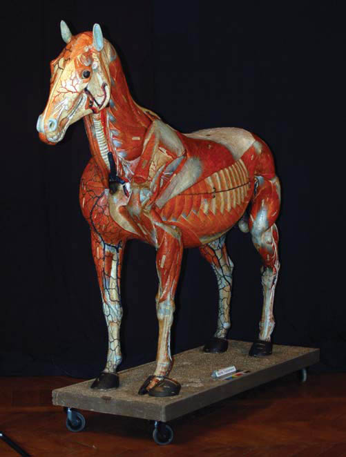 Figure 2b - Left cranio-lateral view of the horse, showing the vessels and deep muscles; the plane of separation of the parts of the trunk can also be seen. |
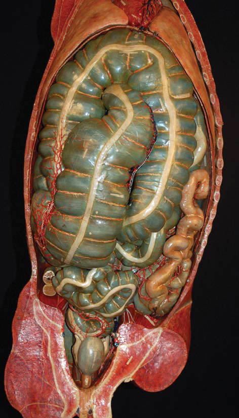 Figure 2c - View of the interior of the trunk after opening, level 1. All the organs are in place and one can see perfectly the folds of the ascending colon and the caecum. |
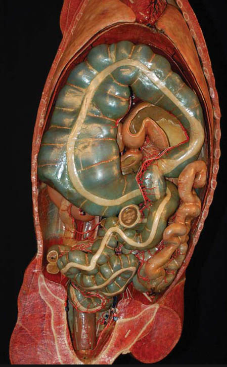 Figure 2d - View of the interior of the trunk after opening, level 2. The ventral part of the ascending colon and the caecum have been removed revealing the dorsal part of the colon and the post-diaphragmatic organs. |
 Figure 2e - View of the interior of the trunk after opening, level 3. The ascending colon and the caecum have been completely removed, revealing the post-diaphragmatic organs and the kidneys. |
Following requests from the cavalry, Auzoux also produced a “cheval incomplet” (literally, “incomplete horse”) designed for the military and the stud farms. This second specimen was the same height and had the same viscera as the “cheval complet”, but the muscles were not detachable; it was made up of 19 parts showing just over half the number of anatomical details, and was accompanied by the same tableau synoptique: (summary chart) the numerals corresponding to the absent anatomical details were simply missing on the specimen (Auzoux, 1855). One of these models, dating from 1846, can be seen at the Fragonard Museum in Maisons-Alfort (just outside Paris), and another in the Science Museum in London. Auzoux even planned to produce three much smaller models, 65 cm in height: one complete, one incomplete and the last simply an écorché (Lequime, 1844), a project of which there is now unfortunately no trace except in the catalogues.
The criticisms of the Royal Academy of Medicine in 1844 were echoed in 1847 by Colonel Jacquemin (Jacquemin, 1847). According to him, the mannequin was “improvable”, a criticism that stung Auzoux, and which spurred him on to produce a new horse, 1.30 m in height and of irreproachable quality, with the profile of a pure-bred Arab steed (Fig. 3).

Figure 3 - A worker poses beside a cheval clastique, type Arab, in the 20th century.
This model was marketed at the beginning of the 1850s10, in two versions, cheval complet and incomplet each accompanied by its own summary chart (Auzoux, 1855).
The other equine models
Auzoux’s next project was to create a series of pathological equine legs complete with various pathologies. Detection of such lesions was of course of the highest importance to horse buyers, whether military or not, but especially to officers of the remonte11. He started with fifty limb models, and added to them over the years (Fig. 4a, b). In this, he was helped and encouraged by his friend Jacquemin who studied and commented on the specimens with great zeal, as is shown by his many letters from the beginning of the 1850s.
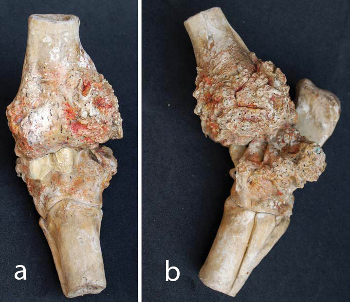
Figure 4 - An example of an osseous lesion (hock) a) anterior; b) lateral view.
Auzoux added a brilliant refinement. As the lesions can be palpated through the skin, he designed some leg models, cut above the hock that were covered with natural skin, placing the student in as realistic a situation as possible. A ‘triptych’ was even successfully marketed, consisting of one dissected limb, one limb affected with bony defects, and a third with soft tissue defects.
Around the same time, Auzoux produced a series of thirty “jaws” (incisor arches) of the horse, copied from natural specimens (Auzoux, 1850 p. 1), and corresponding to the animal’s age12 (Figs. 5a, b). Diagnosing the age of a horse was obviously an important skill for the cavalry officers responsible for procuring horses for the army. Auzoux’s jaws also revealed the tricks that unscrupulous horse dealers would use, such as digging a cavity in the tooth and colouring it with Indian ink make to the animal appear younger.
 Figure 5a - The set of thirty jaws created by Auzoux. |
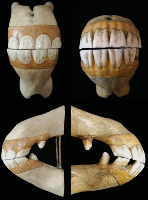 Figure 5b - Detailed views of one of the jaw models. |
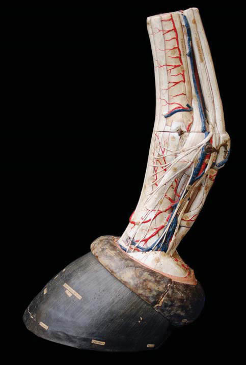
Figure 6 - Model of the foot of the horse.
The hoof is obviously of paramount importance to the well-being and usefulness of the horse. Auzoux made two foot models, one showing the complete anatomy of the region with tendons, ligaments, bones, synovial sheaths, vessels and nerves (Fig. 6),
and the other showing the detailed structure of the hoof (Fig. 7), with the intention of illustrating the theory of the English veterinarian Bracy Clark13, who proposed that the horse’s foot was as deformable as that of other species (Clark, 1817), though this view was ridiculed at the time. Auzoux also produced some isolated horse organs to illustrate his lectures on comparative anatomy and physiology, as well as windowed stomach models (Fig. 8) and the genital organs of the mare.
Marketing and distribution of the models
The complexity of these equine mannequins raised the price considerably, and Auzoux feared that they would suffer a similar fate to that of the Traité de l’anatomie de l’homme by Jean-Baptiste Bourgery14, magnificently – and
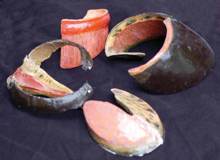
Figure 7 - The separated parts of the hoof, illustrating Bracy Clark’s theory of hoof mechanics.
expensively – illustrated by Nicolas Jacob, but which failed to sell. Auzoux had encountered difficulties trying to sell his homme complet. He had to fight to get the Faculties of Medicine to purchase them, and only managed to achieve his sales targets after persistent reminders and approaches to the public authorities.
Not surprisingly, only the Government departments were able to afford specimens as expensive as Auzoux’s horses. As for the military, the training of the cavalry and artillery was changing, with more emphasis being placed on horsemanship. In 1845, the Minister of War decided to place a cheval clastique at the Royal School of Cavalry in Saumur (Picard, 1890) and in several other depots round the country (Arch. Nat.). At the same time, the Secretary of State for
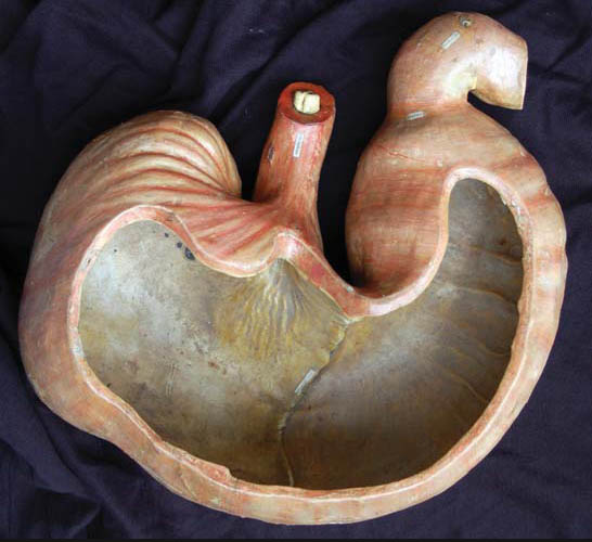
Figure 8 - Windowed stomach of the horse. Marketing and distribution of the models
Commerce and Agriculture started to equip the Veterinary Schools and stud farms with Auzoux’s models. Two years later the Schools of Artillery started to receive the cheval clastique as well as models of the jaws and lesions (Arch. Nat.).
Additional publications: animal husbandry or just advertising?
These successes, however, were not enough to satisfy Auzoux. In a booklet published in 1847 entitled “Of the utility of the anatomie clastique from the point of view of the choice, the employment, and the care of the horse” (Auzoux 1847), he showed how his creations could be used for training the officer cadets, but also – an astonishing generalisation – how they could also be used for the improvement of horse breeding in France. His friend Jacquemin completed the booklet in glowing terms with “An account of the Anatomie clastique of Dr Auzoux and its influence on the training of the cavalry” (Jacquemin, 1847). Other publications with the aim of publicising his creations would soon follow. The bulky and fragile nature of his creations made it difficult to transport them for demonstrations, so these booklets were an important way of reaching his target audience.
Auzoux stressed that in perfecting the training of a cavalry officer, it was necessary to improve not only the quality of the available horses, but also their hygiene and husbandry, although it is not immediately obvious how a cardboard anatomical horse could shed light on the management of contagious diseases! Auzoux had no training in this field, so he made use of his friends and his reading. The archives show that he cut out journal articles, and also borrowed passages from the military training manuals to show how much harm the lack of care had caused during the wars of the Empire (Auzoux, 1847, p. 6). Neglecting the real reason for these problems – the absence of a well-organised system of resupply on top of local shortages – he went on to suggest that with better training, these misfortunes could be avoided.
The issue of improving the national equine herd through improved breeding generated a good deal of controversy in the mid-19th century. Auzoux, (echoing the words of Richard du Cantal) suggested that poor breeding was the result of the ignorance of the stockbreeders (Richard, 1847, p. 404). The introduction of the merino sheep into France in the 18th century was used as an example; this scheme had originally failed due to the lack of understanding about the care that the merino needed. According to Richard du Cantal, to produce horses of good quality, it was necessary first of all to train the breeders in the science of horse husbandry They would then be able to select good stock and to rear them, aware of their needs and ailments.
The dissemination of knowledge in the equestrian world was to become the leitmotiv of Auzoux’s advertising campaign. In 1854, he published the booklet “Insufficiency in France, of the horse for war and for pleasure. Possibility of obtaining them by creating in the Cavalry Regiments, Schools of stock breeders by means of the clastique horse of Doctor Auzoux” (Auzoux, 1854). The idea was that the trained cavalry soldiers would go on to become the apostles of rural horse husbandry…
“The 7 or 8000 released15 each year will go to the centre of horse production that is to say to the farms, taking with them the art of improving the stock.”
The regiments of cavalry and artillery were a captive audience from whom Auzoux hoped to profit. Indeed, it was for them that he had created his cheval incomplet. He envisaged that the military administration would introduce an overall training in horse husbandry for a moderate cost and for the greater good of the French economy. All that was needed was to develop some basic ideas of anatomy, physiology and hygiene that were already being taught as part of the course of military horsemanship, relying on (to make the lessons more expressive)….his cheval clastique (Auzoux, 1847, p. 9). Auzoux expanded on this theme in further booklets dedicated to his collections of lesions and jawbones (Auzoux, 1848, 1853).
Official orders
The results lived up to his expectations. The School of Cavalry of Saumur, flagship of the French Cavalry, started taking his models from July 1845 (Arch. Nat.). In October 1851, they ordered the collection of osseous lesions, the anatomy of the foot, and the ‘Bracy Clark’ hoof (ibid.). On top of that, in 1853 the army purchased “sixty-eight complete copies of the artificial horse”, to be delivered at the rate of twelve per year, as well as a number of jaws and lesions; the regiments of artillery followed suit (ibid.). In military circles, Auzoux had arrived.
In agriculture, the results were, however, less convincing. Auzoux tried hard to get the government departments to adopt the ideas which had been so successful with the military administration. In 1860, he invited the conseils généraux (General Councils) to create a school of agriculture in each département (administrative divisions of France) with the aim of educating all those who were involved in equine husbandry, enticing them by offering a diploma (Auzoux, 1860). The Minister of Agriculture supported his initiative, and in August 1860, sent out a circular letter requesting the Prefects to encourage their Departmental councils to purchase the cheval clastique (Arch. Nat). It was a failure. Only a few models were purchased: the School of Horse-Breeding received one in 184616; the Veterinary School of Alfort, a partner in the project, very soon got its own, which was exchanged for the newer version in June 1856 (Arch. Nat.); this model has since disappeared. They purchased the set of jaws in January 1851 (ibid.). The Veterinary School of Lyon received the jaws at the beginning of 1851, then the cheval complet in December, where it remains to this day.
The equine models, a global success
In the fullness of time, these models went on to become a global success and were distributed all round the world, where they can still be seen today in museums as far afield as New Delhi and Sydney17, proof indeed of their global impact.
Auzoux’s models were such an outstanding success that in 1862, the good doctor was awarded the honour of ‘Officer of the Legion of Honour’, as “inventor of the clastic anatomical appliances used by the army and the military academies” (Le Moniteur, 1862).
Apart from a model of a ruminant stomach, a series of fourteen bovine jaws and two cows’ uteri, (one in a resting state and the other at the end of gestation, with its fetus), Auzoux ventured no further into the anatomy of the domestic animals.
The papier mâché models were quite fragile, and constant use - disassembly and reassembly, inevitably led to damage. There were also considerable losses due to plunder by the Prussian army during the Franco-Prussian war of 1870-1871. So great were the losses that the Minister of War requested from Auzoux in 1874 an estimate for the delivery of “around a hundred and ten cases”, each containing a set of three legs complete with lesions, and the anatomy of the hoof (Arch. Nat.).
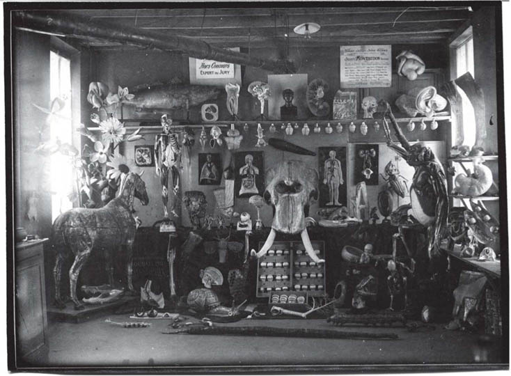
Figure 9. The complete range of Auzoux’s products, c. 1910. On the left is the horse, on the floor just to the right of it are two horse’s legs, one écorché and one complete with skin, the horse’s foot and the Bracy Clark hoof; under the elephant’s head in the centre is the set of horse jaws in their presentation case; on the shelves to the right are the windowed stomach and, above it, another pair of horse’s legs.
It is difficult to put an accurate figure on the scale of Auzoux’s output. While the number of different models was probably between a hundred and a hundred and fifty, the total number of individual parts is probably greater than 300, as certain sets, although indicated by a single catalogue number, were made up of 30-50 parts (Fig. 9). These pieces are today dispersed in many different collections, in very varied conditions. Although well cared-for by collectors and museums, they are unfortunately often neglected in commercial, military or educational collections. In the 19th century, the majority of regiments, colleges and high schools held significant collections of Auzoux’s models. Today, most of these no longer exist or are badly damaged. Conservation of this national treasure should be an urgent priority for France’s cultural institutions (Fig. 10).
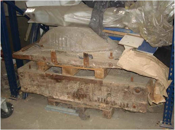
Figure 10. Molds for making the horse models, preserved in Neubourg.
Picture credits
Fig. 1. Archives nationales
Fig. 2a-e. École nationale vétérinaire de Maisons-Alfort.
Figs. 3, 9, 10. Musée de l’écorché d’anatomie du Neubourg.
Figs. 4a, b, 5a, b, 6, 7, 8. Musée de l’École nationale vétérinaire de Maisons-Alfort.
Photography: Figs 1, 2a-e, 3, 7, 8, 9, 10, Degueurce, Christophe. Figs 4, 5, 6, Ruiz, Guillaume.
|
|
Notes:
1Musée de l’écorché d’anatomie, 54 Avenue de la Liberation, 27110 Le Neubourg, France.
2Honoré Fragonard (1732-1799) French doctor, anatomist and veterinarian. Cousin of the painter Jean-Honoré Fragonard. Taught at the first Veterinary School in France (at Maison-Alfort). Some of his preserved specimens can still be seen in the Fragonard Museum near Paris.
3The teenage cadets of the Cavalry School distinguished themselves again in 1940, defending the town during the Battle of Saumur.
4Charles Casimir Beucher, marquis de Saint-Ange (1789-1879)
5A tarred horse écorché can still be seen at the Centre Sportif d’Équitation Militaire de Fontainbleau. As far as we know, it is the last remaining specimen of this type.
6Félix Rigot (1803-1847) appointed Professor of Anatomy at the École royale vétérinaire d’Alfort in 1838. Published a series of papers on the anatomy of the horse.
7Antoine Richard du Cantal (1802 – 1891) French doctor, veterinarian, agronomist and politician. In 1854 he and Geoffroy Saint-Hilaire founded the Zoological Society of Acclimatization.
8Maxime Jacquemin (1795-1863) soldier, scholar and author of works on horse husbandry. Commandant of the Cavalry School of Saumur from 1848.
9The method of fabrication was the same as for the other models. Molds were used to cast the different constituent parts, which were then adjusted, assembled, painted and labelled.
10Auzoux seems not to have publicized this new version very much; perhaps he didn’t want to draw attention to the perceived imperfections of the earlier one. It is therefore difficult to put an exact date on the creation of the new model, which is mentioned only once in the Auzoux archive. An example of the second model can be seen today in the collection of the University of Halle-Wittenberg, Germany.
11Remonte: part of the army responsible for supplying the troops and military establishments with horses.
12Note that only the incisor arches are used to tell the age of a horse.
13Bracy Clark (1771 – 16 December 1860) was an English veterinary surgeon specialising in the horse, who wrote extensively on the structure and functioning of the hoof.
14Jean-Baptiste Marc Bourgery (May 19, 1797 – June 1849) French physician and anatomist. The Traité de l’anatomie de l’homme, published in 8 volumes, is considered to be one of the most comprehensive and beautifully illustrated anatomical works ever published.
15This School, founded by Royal decree in 1840, was notable for its courses in Anatomy, which were supported by the cheval clastique. Auzoux could be forgiven for hoping that each depot and stud farm would be similarly equipped, since teaching had been established in each institution, by ministerial order of June 7th, 1837; one may doubt that this ever happened.
16In the Albert Hall Museum in New Delhi, the anatomical horse “as used by the Cavalry regiments of France” presents, according to the description, “3000 structures on 97 parts”. It is accompanied by a human mannequin, 116 cm in height, and a number of other pieces (flowers, etc.) The Powerhouse in Sydney, Australia, also houses an important collection of Auzoux’s models.
Archives Nationales, 242API (Montandon collection)
Ameline J-F. 1825: Observations sur les pièces d'anatomie de M. le docteur auzoux. Caen, Bonneserre.
Anon. 1843: Article "clastique" in: Anonymous, Dictionnaire de la conversation et de la lecture, t. 14, P., Belin.
Auzoux L. 1825: Notice sur les préparations artificielles de M. Auzoux. Pub., the author.
Auzoux L. 1845: Tableau synoptique du cheval [complet]. Labé.
Auzoux L. 1847: De l'utilité de l'anatomie clastique sous le rapport du choix, de l'emploi, de la conservation du cheval. Pub., the author.
Auzoux L. 1848: Des tares osseuses dans le Cheval. Pub., the author.
Auzoux L. 1850: Mâchoires du cheval et du bœuf. Pub., the author.
Auzoux L. 1854: Insuffisance, en France, du Cheval de Guerre et de luxe. Possibilité de l'obtenir en créant dans les Régiments de cavalerie des Écoles d'éleveurs au moyen du Cheval clastique du Docteur AUZOUX. Firmin-Didot.
Auzoux L. 1855: Tableau synoptique du Cheval [incomplet]. Labé.
Auzoux L. 1858: Leçons élémentaires d'anatomie et de physiologie humaine et comparée. Labé.
Auzoux L. 1860: Insuffisance des Chevaux forts et légers, du Cheval de Guerre et de luxe. Possibilité de l'obtenir en créant dans chaque département des Écoles d'éleveurs. Labé.
Clark B. 1817: Recherches sur la construction du sabot et suite d'expériences sur les effets de la ferrure. Vve Huzard.
Collectif. 1825, 1: Rapport fait par M. Béclard, Duméril, Hippolyte Cloquet, Breschet, Desgenettes sur une pièce d'anatomie artificielle de M. Auzoux, représentant le pied, la jambe, la cuisse et une partie du bassin. Session of 5th November 1823, report published February 1824, in Auzoux, 1825, p. 13-15.
Collectif. 1825, 2: Rapport fait par une commission nommée pour examiner une pièce d'anatomie imitative de M. Auzoux, destinée à représenter la tête, le cou et la partie supérieure du tronc, par M. Worbe, Bégin et Desruelles. In Auzoux, 1825, p. 16-21.
Lafosse PÉ. 1772: Cours d'Hippiatrique. Edme.
Lecoq F. 1843: Traité de l'Extérieur du cheval et des principaux animaux domestiques. Vve Bouchard-Huzard, Lyon, Charles Savy.
Le Moniteur, 16 March 1862, n°75.
Lequime JE. 1844: Exposition des produits de l'industrie nationale en France. Archives de la Médecine belge: 412-415.
Picard L. 1890: Origines de l'École de cavalerie […], Saumur, S. Milon fils, 2 vol.
Renault E. 1845: Rapport fait par M. RENAULT, professeur et directeur de l'École royale vétérinaire d'Alfort, à l'Académie royale de médecine dans sa séance du 22 juillet 1845. Recueil de Médecine Vétérinaire: 843-851.
Richard A (Richard du Cantal). 1847: De la Conformation du Cheval selon les lois de la Physiologie et de la Mécanique. Guiraudet et Jouaust.
Degueurce C. 2012a: Éloge des matières. In: Degueurce C, Delalex H. Beautés intérieures, l'animal à corps ouvert. Réunion des Musées Nationaux, pp. 75-85.
Degueurce C. 2012b: Corps de papier: l'anatomie en papier mâché du Docteur Auzoux. La Martinière.
Degueurce C. 2013 : Les mannequins du Dr Auzoux, une réussite industrielle au service la médecine vétérinaire. Bull Soc Fr Hist Méd Sci Vét 13: 7-33
Dumont B, Dupont A-L, Papillon M-C, Jeannel G-F. 2011: Technical study and conservation of a horse model by Dr Auzoux. Stud Conserv 56: 58-74. https://doi.org/10.1179/sic.2011.56.1.58