Department of Anatomy and Cell Morphology, Faculty of Medicine, University of the Orange Free State, 339 Bloemfontein 9300, Republic of South Africa
Several anatomy departments in South African Medi- cal schools and individuals made different attempts at solv- ing this problem, which were acceptable until plastination appeared on the scene. Most of these applications were in- tended to replace the cartilage. Attempts at our University entailed the use of a section consisting of sternum, inclu- sive of the cartilage, which was covered in plaster of Paris (plus or minus 1cm thickness) and left to dry for a period of 3 months. After removing of the plaster of Paris by tapping the cast with a lightweight object, the thoracic skeleton was then scraped clean and varnished before again being attached to the ribcage. The plaster prevented changing of the shape while drying.
S10; Plastination; Silicone; Cartilage; Thoracic Skeleton;
Willem F. Pretorius, Department of Anatomy and Cell Morphology, Faculty of Medicine, University of the Orange Free State, 339 Bloemfontein 9300, Republic of South Africa

![]()


Historically, serious difficulties were experienced in respect of the preservation of the articulation of the thoracic skeleton, and in particular, the cartilage of the thorax. At- tempts at replacing the cartilage with either rubber plastic or wire and putty produce somewhat inferior imitations.
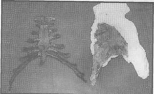
Figure 1. Dried cartilage and plaster of Paris shell
Several anatomy departments in South African Medical schools and individuals made different attempts at solving this problem, which were acceptable until plastination appeared on the scene. Most of these applications were in- tended to replace the cartilage. Attempts at our University entailed the use of a section consisting of sternum, inclusive of the cartilage, which was covered in plaster of Paris (plus or minus 1cm thickness) and left to dry for a period of 3 months. After removing of the plaster of Paris by tapping the cast with a lightweight object, the thoracic skeleton was then scraped clean and varnished before again being attached to the ribcage. The plaster prevented changing of the shape while drying.
Although slightly shrunken this method produced fairly accurate dimensions of the actual structure (figure 1). The only problem being the brittle nature thereof, being at risk of easy breakage if not handled with care.
However, since the introduction of plastination, the following technique is being practiced with great success. The sternum, with cartilage, is removed from a previously embalmed cadaver and plastinated according to the standard S10 technique (von Hagens, 1985). Dehydration is per- formed in cold acetone for 1 month and impregnation for 3 weeks. Care should be taken to secure the structure's dimensions with wire as the cartilage has a tendency to change shape.
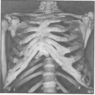
Figure 2. Plastinated cartilage
To great advantage is the fact that it does not produce a bulky mass and has small dimensions. Therefore it dehydrates and impregnates very well and it is a relatively cheap procedure, particularly due to the small mass and dimensions. Cleaning and scraping is done after fast curing. This structure is then attached to the ribs with fishing line.
The final product (figure 2) is exceptionally flexible, and more resistant to breakage. The dimensions are very similar to the actual structure and give the students much better insight into the human anatomy.
Unlike the original or versions previously mentioned which require continuous maintenance, the plastinated specimen can be expected to be maintenance free.
von Hagens G: Heidelberg plastination folder: Collection of all technical leaflets for plastination. Anatomisches Institut 1, Universitat Heidelberg, Heidelberg, Germany 1985.