1Discipline of Clinical Anatomy, Nelson R. Mandela School of Medicine
University of KwaZulu-Natal, South Africa
2Department of Anatomy, School of Medicine, University of Namibia
Windhoek, Namibia
*Dr. Jesse S. Naidu
of blessed memory
Background: This paper reports a descriptive survey carried out to assess the level of awareness of students on the method of plastination and its possible implications in the roll-out and utilization of this learning resource in the Anatomy Department. In view of the recognized challenge emanating from a cadaveric material shortage across anatomy departments in South African Medical Schools, and the unevenness in the bequeathal program across the different regions, it has become imperative to examine other alternatives towards boosting the delivery of the subject of anatomy without undue reliance on cadavers.
Methods: The evaluation data consist of two hundred and sixty-one students across the six disciplines in the Schools of Health Sciences and Laboratory Medicine and Medical Sciences of the University of KwaZulu Natal who volunteered to participate in the survey. Constructs were designed to probe basic bio-geographic data as well as specific aspects relating to plastination with regards to the awareness and possible insights into its relevance or otherwise. Results: This evaluation has identified that the awareness and use of plastinated specimens as an additional learning resource in anatomy remains very low. This was reflected on major probes such as the likelihood to learn the technique (11.88%), if it could replace cadavers (20.99%), if plastinates were better than cadavers (23.75%) and preference to be examined using plastinates (2.3%).
Conclusion: There is low awareness on plastinated specimens and hence greater need for more advocacy on the plastination process to develop the needed passion to embrace the technique by students.
plastination; anatomy education; advocacy
Prof. O. O. Azu, Department of Anatomy, School of Medicine, University of Namibia, Windhoek, Namibia. Tel: +264-612065004, oazu@unam.na; amechi2@yahoo.com

![]()


Research problem statement
The shortage of cadavers for anatomical studies continues to impede teaching and learning of anatomy across the whole spectrum of students in health sciences as well as medicine (Jones, 2002). With increasing numbers of students enrolled in health science faculties and medical schools, the demand for cadaveric materials for dissection has not increased in proportion to the number of students (Azu et al., 2012). The shortage experienced in universities across South Africa necessitates a radical and proactive approach towards revitalizing the ethically preferred body donation pathway (ASSA communique, 2015). Whilst body donation may not be considered appropriate in many South African cultures due to conservative and traditional belief systems, there is an ever-increasing need for a paradigm shift if medical training is to be effective.
Purpose and objectives of study
This study aimed to determine the opinions of undergraduate Health Sciences students about the introduction of plastination and plastinated specimens as an additional resource in the Department of Anatomy, College of Health Sciences, University of KwaZulu-Natal. Our objectives were to assess awareness levels about different anatomical resources and their relevance in the delivery of anatomy teaching and learning across the various disciplines that Anatomy is taught.
In the interim, the onus of addressing the current cadaveric material shortfall lies squarely on medical colleges and anatomy departments nationwide. This has led to the introduction of many innovative mechanisms and processes, including plastination (Cannas and Fuda, 1991; Ravi and Bhat, 2011). While there are extensive debates on the use of cadaveric dissection in medical training (e.g., Furness, 2003; McLachlan et al., 2004), we strongly believe that the experience of studying a real human body in 3-D can never be replaced by computer graphics; it is also important is to learn the technical skills and hand coordination required in the practise of medicine. As Tamura et al. (2014) put it, “understanding the spatial orientation of anatomical structures is critical for learning skills and diagnostic tests”.
Plastination is a unique preservation technique developed by Gunther von Hagens in 1978 (von Hagens et al., 1987). In this method, the water and lipids present in biological tissues are replaced by a polymer such as silicone, epoxy or polyester, which is subsequently hardened, resulting in dry, odorless, and durable specimens. In the nearly four decades since its introduction into the medical terrain, the plastination technique has positively revolutionized the science and art of anatomy. It has brought into question numerous issues of strong ethical and moral dilemma (Jones and Whitaker, 2009). Nonetheless anatomy departments once more have access to a potential ‘goldmine’ of resources for utilization.
The awareness of, and knowledge about, plastinated specimens is important for students, in order to improve their learning in anatomy, but also provides learning experience not available through other instructional delivery systems (Tamura et al., 2014). Anatomy is a traditionally difficult subject for many healthcare students (Johnston, 2010), but is of vital importance as it underpins clinical practice. Hence, the introduction and use of plastinated materials will enable human specimens to be utilized in a fashion like anatomical models with much greater accuracy. Universities across South Africa are experiencing a shortage of cadaveric materials because of an increasing number of medical schools, as well as due to declines in bodies available (ASSA, 2015). There is also no guarantee that the various Anatomy departments would cope with this spiraling surge in the demand for cadavers soon. Therefore, plastination, as a permanent preservation technique of cadaveric materials and body parts, can provide the solution through the production of good quality prosected specimens to be used in teaching and learning. As these plastinates can stand the vagaries of wear and tear, they also offer additional research potential in the field of anatomy. Furthermore, research supports the effectiveness of plastinates in depicting sports injuries, especially for those professionals involved with athletics training and education (Tamura et al., 2014). It may equally be important to assess which teaching/learning resource offers the most benefit to students, bearing in mind that some students may be visual or tactile learners, hence, plastinates may offer unhindered access to additional resource (Sawant and Rizvi, 2015).
The University of KwaZulu-Natal College of Health Sciences undertakes training at undergraduate medical (MBChB) level and offers allied health sciences programs geared towards the promotion of excellence in teaching and research through high quality graduates. The Discipline of Clinical Anatomy runs a functional plastination laboratory located on the Westville Campus of the university, with the aim of producing specimens to add to the available teaching and learning materials in anatomy. In the meantime, there is also the need to understand the dynamics and challenging issues regarding declining anatomy knowledge vis-à-vis curriculum changes globally (reviewed in Papa and Vaccarezza, 2013) and how this will impact on students’ learning outcomes with the introduction of plastinates. The delivery of anatomy subject to medical students at the Nelson R Mandela School of Medicine, University of KwaZulu Natal (UKZN) takes place via a problem-based curriculum, with cadaveric dissection at the center of students’ practical learning. Our study, therefore, investigated the general level of awareness and support for plastination and plastinated specimens in the anatomy repertoire, in consideration of the difficulties. Our future goal is to evaluate the impact of this resource on student performance and through-put as well as how it could support the augmentation of cadaveric material use in the long term.
In this study, a total of 261 survey instruments (questionnaires) were administered to students from seven different disciplines in the School of Health Science and the School of Laboratory Medicine & Medical Science, UKZN, Durban, Westville Campus. The questions were designed to probe the basic demographic data of the participants and their awareness of the plastination process, as well as other constructs to qualitatively assess their use of various learning aids in the department. For example, the first section included a fill-in section for information on basic demographic details like course/degree of study, gender, age, etc. The second section comprised 5 questions regarding the use of teaching aids, types of aids, methods of preserving aids, and reason. The 6th question probed deeper into the various teaching/learning aids used in the department, and participants were to respond by indicating a grading system of 2 (Favorable), 1 (Unfavorable), or 0 (Don’t know) to five different properties of the aid (including handleability, suitability for exams, amongst others). Questions 7 to 16 were designed to gather information relating specifically to plastination: if they have heard of the term, means of awareness, if plastinates are used in the Department, if it is beneficial or better than cadavers, if they will be interested in learning, if they would prefer plastinated specimens for exams. The responses were either a “Yes” or “No”, depending on the nature of the question. As part of the validation process, the questionnaire was pre-tested on 10 students at the Medical School during a dissection hall experience in the discipline.
Ethical considerations
Prior to the administration of the survey instrument, ethical approval was received from the Biomedical Research Ethics Committee (BREC) of the University of KwaZulu-Natal, Durban (BREC no. LMMSEC 025/12). We declared that all the information/data would be handled with confidentiality and would not be used for any other purpose without prior permission from the participants.
The sampling distribution of the questionnaires to various categories of students according to their disciplines/departments in the two schools are shown in Table 1. All the students who volunteered to participate in the survey were given informed consent forms and questionnaires at their lecture venue. The survey instruments were administered by an independent team to maintain confidentiality and reduce bias, after which completed questionnaires were retrieved.
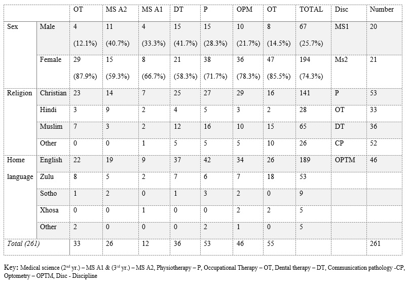 Table 1: Distribution/demographic summary of respondents |
All the data from the questionnaires was collated and analyzed using Microsoft Excel to produce frequency tables. The results are presented as simple descriptive statistics, percentages, tables, and charts.
Inclusion criteria:
Students recruited for the survey included all those who had undertaken courses in Anatomy which included cadaveric dissection in the various programs offered in the Schools of Health Science and Laboratory Medicine and Medical Sciences during the 2012 academic year.
Exclusion criteria:
Students who have not undertaken any course in Anatomy were excluded from the survey.
All 261 questionnaires were returned at the end of the administration (100% response rate). Sixty-seven (25.7%) of the respondents were male, and 194 (74.3%) were female, with a combined age range from 18 to 24 years.
Among the students sampled about their knowledge of plastination, only 84 (32.2%) had heard about it before, 172 (65.9%) had never heard of plastination, and 5 (1.9%) did not respond. We probed further on the use of plastinates, and 48 (18.4%) indicated that they had used plastinated specimens before.
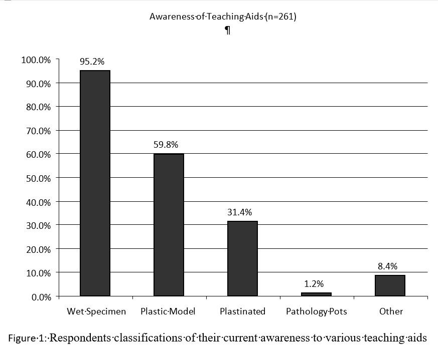
The respondent classifications of current awareness to various teaching aids are illustrated in the bar chart in Figure 1.
Respondents’ opinions on plastinated specimens: 62 (23.7%) said that plastinated specimens are good to use because they are clear and structures can be identified, 17 (6.5%) said plastinates are not better than wet cadavers, and 182 (69.7%) did not respond.
When asked to grade plastinated specimen versus cadaveric material: 21 (8.1%) of respondents said plastinated specimens were better than the wet cadavers, 55 (21.1%) said no, and 185 (70.9%) did not know.
When asked if they would want to learn about the process of plastination: of the 261 respondents, of note is that only 31 (11.9%) said yes, 44 (16.9%) said no, and 186 (71.3%) did not respond.
When asked if plastinated specimens could replace wet cadavers: 54 (21.0%) of the respondents said no while 16 (6.1%) said yes, and 191 (73.2%) did not respond.
On choice of specimens for examination: 33 (12.6%) respondents preferred to be assessed using whole cadaveric specimens, 26 (10.0%) preferred prosected specimens, 10 (3.8%) plastic models, 6 (2.3%) plastinated specimens, no one preferred pathological pots, and 185 (70.88%) did not respond to this question.
In Figures 2 - 6, the following highlights are worth mentioning regarding the grading of the various learning aids used:
Physical appearance/handleability of specimen: only 22.6% (vs 5.7% for cadavers) of respondents felt that plastinates had better physical appearance and handleability (Fig. 2).
Condition of specimen: Most of the students (49.8%) did not favor wet specimens (cadavers) but favored plastic models (56.7%), and the majority (62.8%) did not know about plastinates (Fig. 5).
Suitability for practical examination: most of the respondents (64.0%) did not know that plastinates were suitable for exams, while most of the students (57.9%) favored wet specimens and plastic models (44.8%) for examination; only 23.4% favored plastinated specimens (Fig. 3).
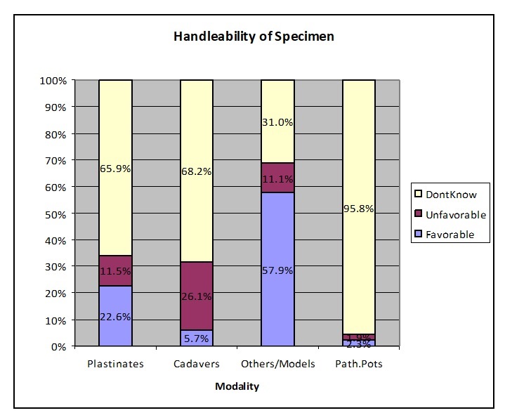 Figure 2: Respondents opinion on handleability of specimens |
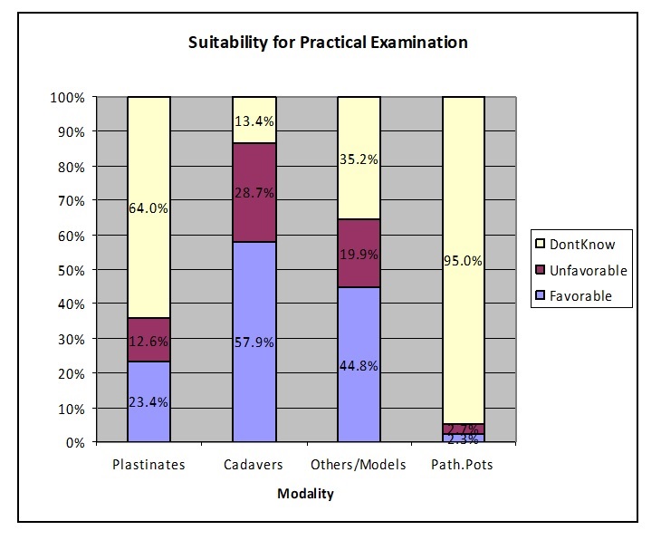 Figure 3: Respondents opinion on suitability of specimens for practical examination |
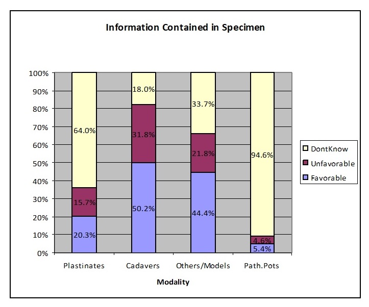 Figure 4: Respondents opinion on information contained in specimens |
 Figure 5: Respondents opinion on condition of specimens |
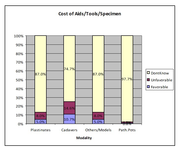 Figure 6: Respondents opinion on cost of specimen |
Information contained in specimens: just over half of the respondents (50.2%) favored wet specimens and plastic models (44.4%), and the majority (64.0%) still did not know about plastinates (Fig. 4).
Cost of aid: the majority of respondents did not know how much the teaching aids cost; this was uniform (Fig. 6).
Plastination, which employs polymers to preserve biological specimens, is recognized as ‘the greatest progress of morphology preservation technology in the 20th century’ (Latorre et al., 2003). The commercial production of plastinates of whole bodies, slices, and animations from plastinates and organs for use in anatomical education are readily available and being produced in various centers across the world where the technology and expertise abound (Azu et al., 2012).
While there is paucity of literature evaluating students’ opinion on plastinated specimens and its efficacy as an adjunct to the repertoire of anatomical teaching and learning aids, there are some reports of positive feedback from use of plastinates by Latorre et al., (2007), Dawson et al., (1990) and Purinton (1991), especially in the developed countries of USA and Europe. However, within the African context, and South Africa in particular, plastination and plastinated specimen use remains poor, perhaps owing to low technological know-how. Many respondents in this report were aware of the various anatomical resources and teaching aids in their learning of anatomy, with cadavers (95.0%), and plastic models (59.8%) being the most widely used. Noticeably, plastinated specimens were less used by the respondents in this study. This shows that the exposure to plastinated specimens in the learning of anatomy at UKZN is still very low, which contrasts with the results of Kamier (2012) where most undergraduates (66.4%) indicated predominant use of plastinated materials, prosected specimens, and models, to augment their learning of anatomy. The minority (33.6%) agreed that the study of anatomy through the dissection of cadavers was the best method of learning anatomy.
Beliefs and religion influence opinions about plastinated specimens and the use of human bodies in learning anatomy. Religious considerations also make it difficult to obtain teaching specimens (Cannas and Fuda, 1991) and enhancing the bequeathal/body donor program. Whilst we did not examine the influence of religion as a factor towards understanding (and/or wanting to learn about plastination and plastinated specimens) by the respondents, literature indicates the strong influence of religion on an individual and how treatment of the dead is handled (whether Islamic, Hindu, Christian, Jewish or Orthodox) (Aramesh, 2009). Though our report shows a high preponderance of Christianity as a religion by most of the respondents, this did not reflect positively in the number who wanted to learn about plastination. There were 54% Christian respondents in the study, but only 12% indicated interest to learn about plastination. We would have expected the latter to be more.
In this study, the respondents declared that they had been exposed primarily to the use of cadavers, prosected specimens and plastic models, which could have contributed to their low awareness of plastinated specimens and plastination. A study by Oyeyipo and Falana (2012) showed that most of the students used cadavers for their study of anatomy (in South-western Nigerian Medical School) and indicated that dissection enhanced their thinking ability. However, this was different from the report of Fruhstorfer et al. (2011) where students (Warwick Medical School, UK) exclusively used plastinated specimens for all regions of the human body, supplemented by non-cadaveric material. Students who no longer have any exposure to wet cadaveric prosections nor the opportunity to participate in cadaveric dissections thus, understandably, think plastinated specimens alone are good. These opposing views and feedback characterize a paradigm shift in the introduction of plastinates into anatomy departments, and could help to inform teachers on how to model the right approach in educating students to use this additional resource. It is interesting to note that the majority of the respondents in this study were females (75%) and a recent report by van der Merwe et al., (2016) shows that over 62% of admissions into the medical schools in South Africa are females, corroborating our report.
The low level of awareness on plastinated specimen usage, particularly as it relates to suitability for teaching purposes as well as its handleability, further highlights part of the challenges faced by experts and users of plastinates. While it enhances visualization of structures, it does not allow for their manipulation, and thus structures such as ligaments and muscles are rendered rigid or semi-rigid (Valdecasas, 2009). Despite this deficiency, the positive aspects and characteristics of plastinates (espoused by Latorre et al., 2007) as a useful teaching tool remains very stimulating and thus can be explored in the local context.
In our study, only 32.98% of the students wanted to learn more about the process of plastination, while 71% did not. This unusual response could be attributed to the fact that many students were not aware of plastination in the first place and therefore had no prior knowledge or entertained serious misconceptions regarding the process. This response was different from a study done by Azu et al. (2012) (carried out with medical students at the University of Uyo-Akwa Ibom State, Nigeria), where over 94% of the students wanted to learn about the technique. Recently, there has also been some doubt amongst scholars in anatomical education about the necessity of using cadaveric material for undergraduate anatomy education (Fruhstorfer et al., 2011). It is argued that not allowing students proper exposure to other methods limits their learning of anatomy. In other words, having limited or no plastinated specimens or other teaching aids, disadvantages students.
As previously mentioned, the fact that most students did not have any experience with plastinated specimens means that they were, therefore, not in a position to judge the value of plastinated specimens. This opens a gap that could be utilized in the introduction of plastinated materials, due to its characteristic nature that favors dry, odorless and tactile nature of specimens. The examination of cadaveric specimens appears to be a crucial element for anatomical studies, and it is therefore questionable to substitute cadaveric learning entirely for contemporary modalities (Fruhstorfer et al., 2011). In this study, the respondents were comfortable with wet cadavers, since this was the only method used in their learning of anatomy and in exams. The respondents graded wet specimens more favorably and suitable for practical examination than plastinated specimens which were graded very low (23.37%).
More than half (57.45%) of the respondents in our study believed that plastinated specimens cannot replace the cadaver and the reason may relate to very low awareness of plastination amongst those sampled, and the fact that they are more exposed to the traditional embalmed cadavers. Plastinated specimens are more often used in the developed countries of Europe and America than in the developing countries of Africa, as illustrated in a study by Fruhstorfer et al. (2011). Their results showed that students highly appreciate plastinated specimens because they were clear and odorless. We previously reported that the advocacy of tissue preservation by plastination has been gradual in developed countries (Azu et al., 2012). The high-cost implications of setting up a plastination laboratory and the necessary technical and human capacity may be responsible for the slow pace of deployment; most developing countries still battle with basic requirements for medical training, with the implication that balancing efficient and effective delivery of anatomy curricula using additional repertoire without compromise on quality will be needed. We believe that the College of Health Sciences at the University of KwaZulu-Natal is yet to benefit from this resource and needs to harness all efforts to aid the learning of anatomy with additional tools like plastinated specimens.
While this study does not champion the complete replacement of cadavers in the training of future medical students, it can point the way forward in resolving the problem of cadaver shortages. Technological developments such as this one are having immense repercussions for clinical anatomy (just as other developments have transformed cell and molecular biology). At the present time, plastinated products not only serve as a training tool, but also as a research tool, and its use is increasing throughout medical schools (Pashaei, 2010).
We expect positive improvements in future anatomy pass rates (seen by students as a difficult subject) with the introduction of plastinates. Another positive outcome of the project is that other clinical departments (like surgery, radiology etc.) in the medical school are expected to benefit from the use of plastinated specimens, which could add to their research capabilities.
A limitation in this study is that it is a descriptive study, which compares teaching and learning aids in the study of anatomy and assesses participants’ subjective opinion regarding the various characteristic of the different aids for teaching and learning anatomy. It is also difficult to extrapolate our findings to a larger student cohort in view of the limited number sampled, and hence, future studies should incorporate this factor.
We conclude that the use of plastination as a cadaveric preservation method is not widely known by students, and the use of plastinated specimens is still very low at the University of KwaZulu-Natal, Durban. We believe that the introduction of plastinated specimens will improve student learning in anatomy, especially as cadavers are becoming scarce. Since plastinated specimens can be used repeatedly with damage less to the material, this can help to augment the number of specimens available to students who enroll for anatomy.
Conflict of interest: None declared by the authors.
Acknowledgements: Ms. Nosipho Nzimande for her assistance and support and Dr. Jacqueline van Wyk for her suggestions on the survey instruments.
Aramesh K. 2009: The ownership of human body: An Islamic perspective. J Med Ethics Hist Med 2:4.
ASSA communique. 2015: http://www.sabc.co.za/news/a/c15ba30049787268a0cda1623266b54e/Shortage-of-cadavers-at-SA-universities-20151408 Accessed 26th October 2015.
Azu OO, Peter AI, Etuknwa BT, Ekandem GJ. 2012: The awareness of medical students in Nigerian universities about the use of plastinated specimens for anatomical studies. Maced J Med Sci 5: 5-9.
https://doi.org/10.3889/MJMS.1857-5773.2011.0202
Cannas M, Fuda P. 1991: Plastination of old formalin-fixed species. J Int Soc Plastination 5: 11-15.
https://doi.org/10.56507/IYFR4714
Dawson TP, James RS, Williams GT. 1990: Silicone plastinated pathology specimens and their teaching potential. J Pathol 162:265-272.
https://doi.org/10.1002/path.1711620314
Fruhstorfer BH, Palmer J, Brydges S, Abrahams PH. 2011: The use of plastinated prosections for teaching anatomy - the view of medical students on the value of this learning resource. Clin Anat 24: 246-252.
https://doi.org/10.1002/ca.21107
Furness P. 2003: Consent to using human tissue. BMJ 327: 759-60.
https://doi.org/10.1136/bmj.327.7418.759
Jones DG. 2002: Re-inventing anatomy: The impact of plastination on how we see the human body. Clin Anat 15:436-440.
https://doi.org/10.1002/ca.10040
Jones DG, Whitaker MI. 2009: Engaging with plastination and the Body Worlds phenomenon: A cultural and intellectual challenge for anatomists. Clin Anat 22: 770-776.
https://doi.org/10.1002/ca.20824
Johnston, ANB. 2010: Anatomy for nurses: providing students with the best learning experience. Nurse Educ Pract 10: 222-226.
https://doi.org/10.1016/j.nepr.2009.11.009
Kamier MA. 2012: Attitude and views of medical students toward anatomy learnt in the preclinical phase at King Khalid University. J Fam Community Med 19: 190-193.
https://doi.org/10.4103/2230-8229.102320
Latorre RM, Garcia-Sanz MP, Moreno M, Hernandez F, Gil F, Lopez O, Ayala MD, Ramirez G, Vazquez JM, Arencibia A, Henry RW. 2007: How useful is plastination in learning anatomy? J Vet Med Educ 34: 172-176.
https://doi.org/10.3138/jvme.34.2.172
Lattore R, Arencibia A, Gil F, Rivero M, Rameriz G, Vaquezauton JM, Henry RW. 2003: Plastinated slices: An aid to interpreting MR Images of the equine tarsus. J Int Soc Plastination 18: 14-22.
https://doi.org/10.56507/KDJG6154
McLachlan JC, Bligh J, Bradley P, Searle J. 2004: Teaching anatomy without cadavers. Med Educ 38: 418-24.
https://doi.org/10.1046/j.1365-2923.2004.01795.x
Oyeyipo IP, Falana BA. 2012: Attitude of preclinical students to cadaver dissection in a South West Nigerian medical school. Int J Trop Med 7: 1-5.
https://doi.org/10.3923/ijtmed.2012.1.5
Papa V, Vaccarezza M. 2013: Teaching anatomy in the XXI century: New aspects and pitfalls. Sci World J 2013: Article ID 310348, 5 pages
https://doi.org/10.1155/2013/310348
Pashaei S. 2010: A brief review on the history, methods, and applications of plastination. Int J Morphol 28: 1075-1079.
https://doi.org/10.4067/S0717-95022010000400014
Purinton P. 1991: Plastinated brains used with computer assisted learning modules for teaching veterinary neuroanatomy laboratories. J Int Soc Plastination 5:16-19.
https://doi.org/10.56507/WDQQ3260
Sawant SP, Rizvi S. 2015: Teaching anatomy to undergraduate students. Int J Anat Res 3:1212-15.
https://doi.org/10.16965/ijar.2015.172
Spoorthi Banavar Ravi, Vidya Manohar Bhat. 2011: Plastination: A novel, innovative teaching adjunct in oral pathology. J Oral Maxillofac Pathol 15: 133-137.
https://doi.org/10.4103/0973-029X.84475
Tamura K, Stickley CD, Labrash SJ, Lozanoff S. 2014: Effectiveness of plastinated anatomical specimens depicting common sports injuries to enhance musculoskeletal injury evaluation education. Athl Train Educ J 9: 174-181.
https://doi.org/10.4085/0904174
Valdecasas A. 2009: Understanding complex systems: lessons from Auzoux's and von Hagens's anatomical models. J Biosci 34:835-843.
https://doi.org/10.1007/s12038-009-0097-0
van der Merwe LJ, van Zyl GJ, St Clair Gibson A, Viljoen M, Iputo JE, Mammen M, Chitha W, Perez AM, Hartman N, Fonn S, Green-Thompson L, Ayo-Ysuf OA, Botha GC, Manning D, Botha SJ, Hift R, Retief P, van Heerden BB, Volmink J. 2016: South African medical schools: Current state of selection criteria and medical students' demographic profile. S Afr Med J 106:76-81.
https://doi.org/10.7196/SAMJ.2016.v106i1.9913
von Hagens G, Tiedemann K and Kriz W. 1987: The current potential of plastination. Anat Embryol 175: 411-421.
https://doi.org/10.1007/BF00309677