1Department of Anatomy, Dalian Medical University, Dalian, P. R. China
2Dalian Hoffen Bio-Technique Co. Ltd., Dalian
Plastination is a method of preserving biological tissues with a curable polymer. Sheet plastination is a method of preparing plastinated tissue slices for education and research. Both epoxy and polyester sheet plastination are currently used. P45 sheet plastination produces an intact, semi-transparent anatomical structure, with well highlighted connective tissue. There is little information in the literature regarding the physical properties of P45 plastinated specimens. Shrinkage during plastination is to be expected. In this study we present data on the shrinkage of the following P45 plastinated tissue slices: eight head and neck sections, five thoracic sections, eight abdominal sections, five pelvic sections, and three arm sections. The standard P45 protocol was followed, and a digital image of the specimens was taken before and after the plastination process. Analysis of the images showed that shrinkage varied between 6.39 (±3.9) % for cerebral cortex, and 19.86 (±1.68) % for lung tissue.
sheet plastination; polyester method; body slices; polyester resin; P45 technique; tissue shrinkage
Sui Hong-Jin. Tel.: +8613904287577; Email: suihj@hotmail.com

![]()


Plastination is simply the process of substituting water and lipid molecules from biological tissues with curable polymer. It is considered a major improvement in the preservation of biological specimens (Riederer, 2014; McRae et al., 2015). Plastinated specimens pose no health hazards (Henry et al., 1997; Sivrev, 2012).
Sheet plastination has been used over the decades as a teaching and research tool. In teaching, for instance, the study of topographical anatomy is helpful for interpreting MRI and CT images (Thomas, 2004), and slice plastinates aid in the appreciation and understanding of sectional anatomy in biomedical images. Sheet plastination also plays essential roles in clinical anatomy research, it has been used in the study of joints, body cavities and spaces, bones, neurovascular structures, body ligaments, muscles, organs, and 3D computational reconstruction of anatomical structures (Sora & Genser-Strobl, 2007). The usage of sheet plastinated specimens in teaching and research is due to the fact that sheet plastination presents the body structures in a non-collapsed and non-dislocated form (Sora et al., 2002). P45 sheet plastination is a polyester resin plastination method used to preserve biological tissues. P45 plastinated sheets are semi-transparent slice sections, with the internal structures of the specimen clearly revealed (Gao et al., 2006).
Shrinkage of biological specimens during plastination is expected. The shrinkage values can be used to validate both morphometric and 3D reconstruction measurements. This study addresses tissue shrinkage after P45 sheet plastination.
Two cadaveric specimens were used for this study, and approval for the study was given by the Department of Anatomy, Dalian Medical University. The following sections were obtained from the specimens: eight head and neck sections, five thoracic sections, eight abdominal sections, five pelvic sections, and three arm sections.
P45 sheet plastination procedure
A detailed P45 sheet plastination procedure has been documented by Sui and Henry (2007), and Okoye and Sui (2019). The cadavers were formalin-fixed prior to processing. The specimens were first frozen in an ultra-cold deep freezer at -70° C for about two weeks.
Slicing
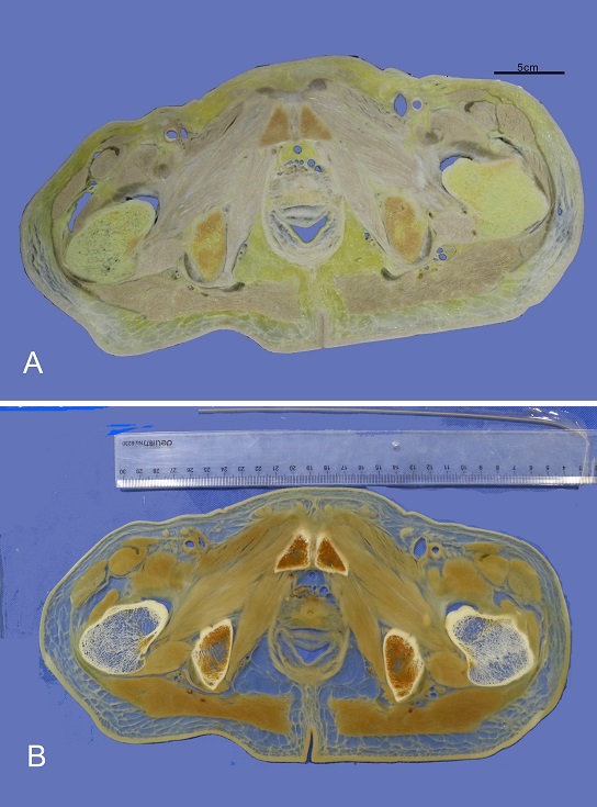 Figure 1. Pelvic section before the P45 plastination process (A) and after P45 plastination (B). |
The specimens were embedded in polyurethane and then sectioned using a bandsaw with a blade thickness of 0.3 mm, and a cutting speed of 40 m/s. The section thickness was 2 mm. Between adjacent slices, tissue lost as tissue dust while sawing was approximately 1 mm thick (Fig. 1a).
The sliced body sections were placed on polyethylene grid with a cotton fibre screen. The sawdust on the sliced sections was removed with small stream of gently running water. The grids with their washed slices were then stacked and tied with twine to hold each stack as a unit. The stacked units were then transferred into the first cold acetone (-25° C) bath
Bleaching
The stacked units were immersed in 5% hydrogen peroxide overnight to improve tissue color brightness and transparency. Subsequently, excess bleach was washed off by washing the slices in running water for one hour or more.
Dehydration
The stacked units of slices were firstly precooled to 5° C in order to avoid the formation of ice crystals and shrinkage, before being submerged in a 100% acetone bath at -25° C for one week. The stacked units were then transferred into another fresh acetone bath at -15° C for one week. Finally, the stacked units were transferred into 100% acetone at room temperature for another week.
Preparing the P45 polyester mixture
The impregnation resin mixture was prepared as follows: 1000 ml P45 resin (Hoffen polyester, China) was mixed with 10 g of P45A, 30 ml P45B and 5 g of P45C. P45A and P45C are plasticizers and P45B is a hardener. The open vertical chamber was then prepared.
Building the casting chamber
The casting chamber used in this technique is an open vertical chamber. The top part of the chamber is left open, while the bottom and two sides of the casting chamber are clamped. The open vertical chamber was placed vertically in the vacuum chamber, i.e. standing on its bottom end. The chamber was constructed from two plates of 5 mm tempered glass, flexible 4 mm latex tubing, and several large fold-back clamps.
The tubing was intercalated between the two tempered glass sheets, and the glass and tubing were then clamped together using fold back clamps on three sides, leaving the top open.
Forced impregnation of the slices
The body sections were removed from the final acetone bath and placed into the open vertical chambers. The chambers were then filled with P45 polyester resin mixture (Hoffen polyester, China) using a customized funnel at the top open part of the chamber.
The vertical chambers, with the top part are left open, were then placed upright into a vacuum chamber about one metre deep. The vacuum chamber was sealed, and the absolute pressure was progressively decreased to 20 mm Hg, 10 mm Hg, 5 mm Hg, and 0 mm Hg, maintaining slow bubble production and release. The pressure was maintained at 0 mm Hg until bubbling ceased. The impregnation process was performed at room temperature and was completed in approximately 8 hours.
Curing
After impregnation, the open vertical chambers were transferred to a water bath at 40° C for curing, for three days.
Finishing
After curing, the open vertical chambers were removed from the water bath and dismantled. The P45 slice were wrapped with a plastic sheet or light-weight foil.
 Figure 2. A thoracic section uploaded into the Image J software before the plastination process (A), and after P45 plastination (B). The dotted line is the tracing line used by the software for measuring the superior surface area of the organ |
Measurement of the tissues
A calibrated photographic documentation of the slice sections was taken before tissue slicing and after P45 plastination (i.e. after curing) (Fig. 1b). The photographs were uploaded to Image J software (Image J 1.52i), and the surface area of each organ was measured (Fig. 2).
The brain, kidney, liver, muscles and spleen were measured. The muscles measured were the gluteus maximus and the triceps. The cerebral cortex of the brain was measured on the brain sections. Measurements of each organ before slicing and after curing were documented, and the percentage shrinkage was calculated.
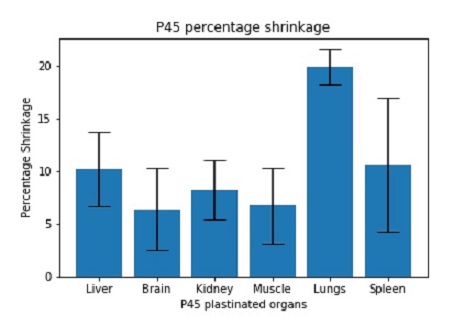 Figure 3. Graphical representation of the percentage tissue shrinkage |
The P45 sections were in good condition, the slices were semi-transparent and the connective tissues and organs in each sheet were all intact. The measurements of the organs before and after P45 plastination, and the percentage shrinkage of the tissues are presented in Table 1.
The lung tissue showed the highest mean percentage shrinkage value, and the brain tissue the lowest mean percentage shrinkage (Fig. 3).
| Liver | Brain | Kidney | Muscle | Lung | Spleen | ||||
| No. of slices measured | 5 | 8 | 5 | 5 | 5 | 5 | |||
| Area of superior surface before plastination (cm2) | 493.92 | 524.98 | 83.38 | 61.21 | 453.890 | 143.21 | |||
| Area of superior surface after plastination (cm2) | 443.59 | 491.40 | 76.49 | 57.09 | 363.75 | 127.96 | |||
| Average percentage (%) shrinkage SD |
3.5 |
6.39
3.9 |
8.26
2.8 |
|
19.86
1.68 |
10.64
6.35 |
Table 1 Surface area and average shrinkage of different tissues before and after P45 sheet plastination.
Shrinkage leads to the reduction of the actual measurement of the tissue. Thus, the organ shrinkage value is useful in determining accurate dimensions of plastinated specimens especially in morphometric measurements and 3D image reconstructions.
Studies on the shrinkage of thin E12 and standard E12 body sections and also P40 brain slices have been reported. While E12 and P40 are popular sheet plastination techniques, the P45 technique is not currently widely used. However, the shrinkage of this technique has until now not been reported (Sora et al., 2002; Sora et al., 2015).
Several factors can affect tissue shrinkage. Two factors have been strongly advocated:
(1) The dehydrating temperature affects shrinkage, though dehydrating at low temperatures (-25° to +5°C) will reduce shrinkage (von Hagens, 1985, Brown et al., 2002), though the tissue transparency will be negatively affected because of minimal defatting (Cook and Al-Ali, 1997). This can be tackled by increasing the degreasing time (Sora et al., 2002), but this might also impact on the shrinkage of the processed tissue.
(2) The shrinkage of the impregnating resin itself may also affect tissue shrinkage during plastination.
In this study, the sections were measured only before and after P45 plastination, and did not include shrinkage during dehydration or impregnation. Taking the specimens out for measurement during the plastination process can impact negatively on the shrinkage of the plastinated section.
The measurement carried out was a bi-dimensional measurement of the length & width of the organs. The different P45 organ sections had different shrinkage rates. The mean percentage reduction of the brain, kidney and muscles in this study were below 10%, while the spleen, lungs and liver were above 10%. As expected, the lung shrinkage was greater than 10%. The shrinkage of the lung sections was maximal, while the shrinkage of the brain sections was minimal. This may be attributed to the properties of the organs – though the brain is soft, its cells are densely packed. The lungs, on the other hand, are spongy and have certain elastic properties. Thus, in addition to dehydration and shrinkage of the resin, the properties of the organ or tissue being plastinated may impact its shrinkage.
The percentage shrinkage of brain specimens in this technique was similar to that found in the P40 technique for brain tissue (Sora et al., 1999) but differs for kidneys (Pereira-Sampaio et al., 2011). In the P45 technique, the bleaching procedure is performed before dehydration, while in the P40 process, dehydration is performed before bleaching. The duration of plastination using polyester resin (P40, P45) is relatively short when compared to their epoxy counterpart. However, P45 requires no UV light for curing, unlike the P40 technique.
Morphometric and 3D reconstruction measurements from P45 sections should take into consideration the shrinkage of the tissues, and also the tissue loss while sawing the specimens. The values ascertained in the present study can provide a useful estimate of shrinkage in future studies.
Brown MA, Reed RB, Henry RW. 2002: Effects of dehydration mediums and temperature on total dehydration time and tissue shrinkage. J Int Soc Plastination 17:28-33.
https://doi.org/10.56507/XNQM4606
Cook P, Al-Ali S. 1997: Submacroscopic interpretation of human sectional anatomy using plastinated E12 sections. J Int Soc Plastination 12(2):17-27.
https://doi.org/10.56507/XICY2283
Gao H, Liu J, Yu S, Sui H. 2006: A new polyester technique for sheet plastination. J Int Soc Plastination 21:7-10.
https://doi.org/10.56507/JPVW6850
Henry RW, Janick L, Henry C. 1997: Specimen preparation for silicone plastination. J Int Soc Plastination 12(1):13-7.
https://doi.org/10.56507/HVSK9838
McRae KE, Davies G, Easteal R, Smith GN. 2015: Creation of plastinated placentas as a novel teaching resource for medical education in obstetrics and gynaecology. Placenta 36:1045-1051.
https://doi.org/10.1016/j.placenta.2015.06.018
Okoye CS, Sui H-J. 2019: Updated protocol of Hoffen P45 sheet plastination technique. J Plastination 31(2).
https://doi.org/10.56507/KDAS3395
Pereira-Sampaio MA, Marques-Sampaio BP, Sampaio FJ, Henry RW. 2011: Shrinkage of renal tissue after impregnation via the cold Biodur plastination technique. Anat Rec 294:1418-1422.
https://doi.org/10.1002/ar.21432
Riederer BM. 2014: Plastination and its importance in teaching anatomy. Critical points for long-term preservation of human tissue. J Anat 224(3):309-315.
https://doi.org/10.1111/joa.12056
Sora MC, Brugger P, Traxler H. 1999: P40 Plastination of human brain slices: comparison between different immersion and impregnation conditions. J Int Soc Plastination 14(1): 22-24.
https://doi.org/10.56507/XLSJ5724
Sora MC, Brugger PC, Strobl B. 2002: Shrinkage during E12 Plastination. J Int Soc Plastination 17:23-27.
https://doi.org/10.56507/DIUH4490
Sora MC, Genser-Strobl B. 2007: The sectional anatomy of the carpal tunnel and its related neurovascular structures studied by using plastination. Eur J Neurol 12(5):380-384
https://doi.org/10.1111/j.1468-1331.2004.01034.x
Sora MC, Binder M, Matusz P, Ples H, Sas I. 2015: Slice plastination and shrinkage. Mater Plast 52(2):186-189.
Sui HJ, Henry RW. 2007: Polyester plastination of biological tissue: Hoffen P45 technique. J Int Soc Plastination 22:78-81.
https://doi.org/10.56507/IJSL3572
Sivrev, D. 2012: Safety and durable P35 and P40 plastination slices of anatomical objects. Conference: actual questions of theoretical and practical medicine. Nalchik, Russia 75:109-111.
Thomas M, Steinke H. 2004: Thin-layer plastination of the shoulder. CSMI 1:9-14