Neurosurgery, Medical School, University of Mainz, Mainz, GERMANY
Neuroanatomy, studied via the endoscope on plastinated specimens, will play a key role in endoscopial neurosurgery in the future. Plastinated specimens are proving to be an invaluable aid for training in all phases of the neurosciences.
Silicone; S10; Biodur
D. M. Resch, Neurosurgery, Medical School, University of Mainz, Mainz, GERMANY

![]()


At the neurosurgical clinic, University of Mainz, plastinated specimens have been prepared to help establish endoscopial neuroanatomy and plastinated crania are now used for neuroendoscopial training. Plastinated specimens are dry and available for use in any environment; as opposed to wet, toxic formalin- fixed specimens which are not practical for such training (Resch, 1989). Plastinated specimens reveal precise anatomical detail {Resch and Perneczky, 1990). As the endoscope is manipulated through the prosected regions of the plastinated specimen, new views are possible with the plastinated specimens. Hand-eye coordination is enhanced by using this set up.
Amputated, formalin-fixed crania were prosected through a dissection microscope. Keyhole approaches up to 15 cm deep were prepared to demonstrate selected areas of neuroanatomy. Four specimens were prepared and were plastinated using freeze substitution and the standard S 10 method for impregnation of the silicone polymer (von Hagens, 1985).
The vessels were injected with colored PEM prior to amputation. The specimens were immersion fixed in a 10% formaldehyde solution with perfusion of the ventricles and subarachnoid space, after which they were dissected. The specimens were dehydrated by freeze substitution in preparation for plastination. The standard S10/S3 technique was used to produce the plastinated specimens (von Hagens, 1985).
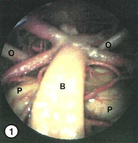 Figure 1. The entire transoral aspect of the brain stem of the plastinated specimen may observed and magnified using the endoscope. The basilar artery (B) may be followed along the pons (P) to its quatrification. Both oculomotor nerves (0) are in focus. |
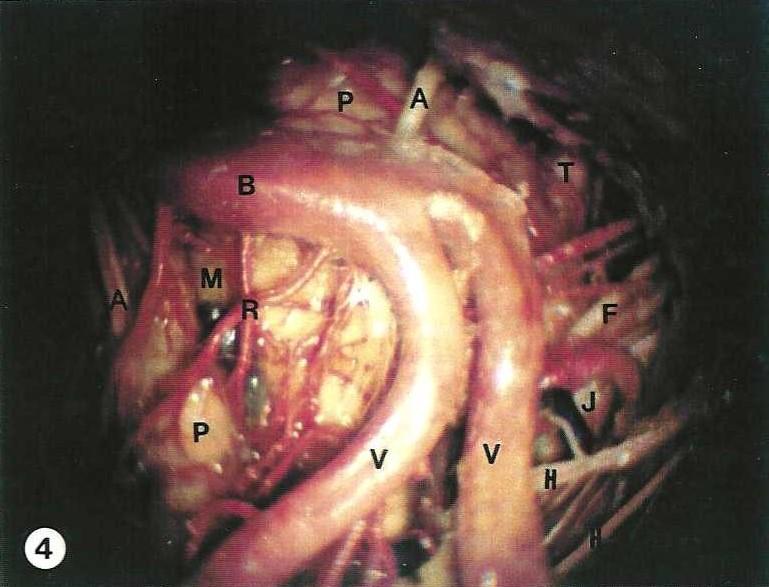 Figure 4. Endoscopic view of Figure 2, the transoral aspect of the brain stem. The optical characteristics are a typical "fish eye" view yielding a much broader field. Many nerves can be followed to their foramina, Abducens (A), Acoustic and Facial (F), Hypoglossal (H), the Jugular foramen group (J) and Trigeminal (T). Basilar artery (B), Basilar vein (M), Pons (P), Pontine perforators (R), Vertebral arteries (V). |
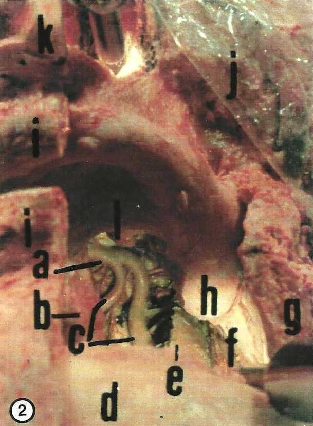 Figure 2. Transoral view of the brain stem through a large window in a plastinated specimen. Alveolar process (h), Basilar artery (a), Hard palate (I), Mandible (d), Medulla oblongata (origin of hypoglossal nerve) (k), Pontomedullary sulcus (j), Soft tissue of the face (i, g, f), Tongue (b), Vertebral artery (c), Vertebral joint (e). |
|
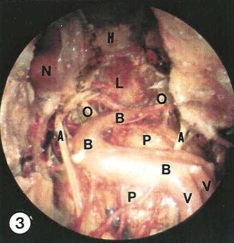 Figure 3. In the plastinated specimen the endoscope may be placed at different depths and the syntopy of previously hidden structures can be studied. Behind the base of the cranium in the sphenoidal sinus the impression of the carotid artery (N) is seen. Abducens nerve (A), Basilar artery (B), Brain stem structures [Hypophysis (H), Hypothalamus (L), Pons (P)], Oculomotor nerve (0), Vertebral arteries (V). |
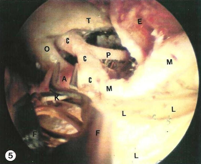 Figure 5. Through a right frontotemporal approach, in the plastinated specimen using the endoscope, an aneurysm clip (K) is introduced with a clip forceps (F) during a training session. Anterior cerebral artery (A), Carotid artery (C), Frontal lobe (L), Middle cerebral artery (M), Optic nerve (0), Posterior communicating artery (P), Temporal lobe (E), Tentorial notch (T). |
Resch, KDM: Plastinated specimens for demonstration of microsurgical approaches to the base of the cranium. J Int Soc Plastination 3:29- 33, 1989.
https://doi.org/10.56507/FUGF5217
Resch, KDM, A Perneczky: The use of plastinated specimens in planning microsurgical approaches to the skull and brain base. Presented at The 5th International Conference on Plastination, Faculty of Medicine, University of Heidelberg, Germany July 1990 J Int Soc Plastination 4:12, 1990.
von Hagens G: Heidelberg Plastination Folder: Collection of all technical leaflets for plastination. Anatomisches Institut 1, Universitat Heidelberg, 1985.