Departement de chimie-biologie, Universite du Quebec a Trois-Rivieres, Trois-Rivieres, Quebec, Canada
In the last centuries, teaching of anatomy required anatomical models because of the lack of satisfactory preservation techniques. These models had to meet many requirements: they should be as realistic as possible, not too delicate, contain some movable parts, and not be too expensive. Many different kinds of materials have been used in that aim. Nowadays, most of these models are rightly regarded as masterpieces which paved the way for current anatomical teaching. One of their major defaults was the lack of authenticity: in some ways, this last step was cleared by the invention of plastination some twenty years ago.
Anatomical models, Felice Fontana, Angelique Du Coudray, Louis Auzoux
Dr. R. Olry, Departement de chimie-biologie, University du Quebec a Trois-Rivieres, C.P. 500, Trois-Rivieres, Quebec, CanadaG9A5H7. Telephone: 819 376 5053 /Fax: 819 3765084. Email: Regis_Olry@uqtr.uquebec.ca

![]()


For about twenty years, plastinated specimens are rightly regarded as powerful teaching aids in human and comparative anatomy (see Grondin and Olry, 1996, for review). However, the use of anatomical models for the education of students (medicine, surgery, midwifery, fine arts) probably goes back to many centuries. Though dissection was aknowledged for a long time as the best way to understand anatomy, the study and teaching of the human anatomy came up against two real problems in the course of their history: the ban of the dissecting human bodies in the Middle Ages on the one hand, and the lack of bodies in the 17th and 18th centuries on the other hand (Olry, 1999). It was therefore necessary to show ingenuity to bypass these problems: for want of human specimens, it became necessary to make anatomical models. All kinds of materials have been used, more or less successfully: wax, wood, ivory, cardboard, bronze, fabric, plaster, rubber, and plastic. This paper summarizes the history of these models of artificial anatomy. Anatomical specimens, such as the medical mummies of the Burns collection (Wade, 1998) or the ghostly specimens by Honore Fragonard (Hugues, 1990) will not be described in this paper.
Wax anatomical models
Wax anatomical models are probably the most striking masterpieces of artificial anatomy (Le Minor and Puygrenier, 1989; Lemire, 1990). Ceroplasty spread in the 17th and 18th centuries, mainly in Italy and France. The most famous artists were Gaetano Giulio Zummo (1656-1701), Giovanni Manzolini (1700-1755), Ercole Lelli (1702-1766), Anna Morandi (1716-1774), Felice Fontana (1730-1805), Clemente Susini (1754-1814), Francesco Calenzuoli (1796-1829), Luigi Calamai (1800-1851), Giovanni Lusini (1809-1889) and Egisto Tortori (1829-1893) in Italy; Antoine Benoist (1629- 1717), Guillaume Desnoues (ca. 1650-ca. 1735), Marie- Catherine Biheron (1719-1796), Guillaume Curtius (1737-1794), Andre-Pierre Pinson (1746-?), and Jean-Baptiste Laumonier (1749-1818) in France.
Fabrication of wax anatomical models usually required a close collaboration of an anatomist (or a surgeon) and a sculptor. The first step was the dissection of the regions or viscera to be casted. Usually, four to five anatomical specimens were necessary to display the complex regions, such as the head. The second step was the making of a plaster cast. The surface of the specimen was first coated with fat so that the plaster does not stick, and a thin coat of liquid plaster was then poured on the specimen. The cast was often removed in pieces: some threads placed on the specimen made the breaking up of the cast easier. The third step was the making of the wax model. The plaster cast was coated with liquid soap or walnut oil in order to block up the pore of the plaster.
Wax (Smyrne white wax, beeswax, " spermaceti" obtained from the head of sperm whale) and additives (mutton suet, olive oil, Venice turpentine) were then applied by thin successive coats. The fourth and last step aimed at assembling the different components of the model, correcting the defects due to the removal, painting or sticking the superficial vessels and nerves, and finally glazing the model.
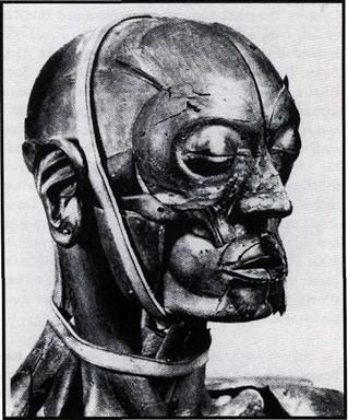
Figure 1. Wooden anatomical model by Felice Fontana (detail, taken from Lemire, 1990).
Wooden anatomical models
Egyptians are known to have produced human models made of wood and sometimes covered with painted plaster. Some of these models have come down to our own day. However, it seems that these models never had any educational purpose. According to Herodote, they were used on the occasion of banquets, and the host displayed them to the guests and said: " Look at this man; you will look like him after your death; so drink now, and enjoy yourself " (Herodote, cited in Lauth, 1815).
The most famous anatomical wooden models were made by the Italian abbot Felice Fontana in the late eighteenth century (figure 1). One of his masterpieces was a life-size standing man, composed of 3000 movable parts, each of them showing about 100 details! (Fontana, 1795). This male model, a painstaking task which took about 10 years, could even be converted into a female body. In 1796, general Bonaparte was so impressed that he ordered a copy of this model, but its manufacture was delayed by the vicissitudes of the war. In 1805, the interest had fallen off: " the wooden specimen costed 30.000 liras and was in fact not a very satisfactory work and had not the least usefulness " (Sonolet, 1975).

Figure 2. Ivory anatomical models (France or Italy, early 18th century), probably for the instruction of midwives (Collection Librairie Alain Brieux, 1986, with kind permission).
Ivory anatomical models for the instruction of midwives
The most beautiful anatomical manikins in ivory were produced in France and Italy in the seventeenth and eighteenth centuries. Some ivory models were produced to show special parts of the human body: a model of the ear, attributed to the Venetian artisan Giambattista Verle and probably inspired by Valsalva's treatise on the human ear (Valsalva, 1735), is currently kept in the Museum of La Specola at Florence. However, most of the ivory anatomical models displayed the whole female body, often pregnant, and contained movable parts (abdominal wall, uterus with foetus, intestines) (figure 2). There is some doubt as to the exact purpose for which they were used, but it is most probable that they were for the instruction of midwifes (Weil, no date; Brieux, 1986). Whatever the materials used, numerous midwives are known to have used anatomical models of pregnant women for the instruction of their pupils or colleagues. Among them, one of the most famous was Angelique Du Coudray (1712-1789) (figure 3). She taught in France and The Netherlands with the aid of a manikin which she had made herself: " I made up my

Figure 3. Portrait of Angelique Du Coudray (1712-1789) (taken from Saint-Restitut, 1989).
mind to make my lessons palpable (by mean of) a machine which I made in that aim, which represented the pelvis of a woman, the uterus..., its ligaments..., the umbilical cord composed of two arteries and one vein..., a model of a life-size child (who could be placed) in different positions " (Saint-Restitut, 1989).
Louis Auzoux and his clastic anatomy
Louis-Thomas-Jerome Auzoux, the son of Norman farmers, was born on April 7, 1797, in Saint-Aubin d'Ecrosville, France (Hirsch, 1888; Saint-Restitut, 1986) (figure 4). He studied medicine in Paris, under Guillaume Dupuy tren at the Hotel-Dieu (1818), and subsequently under Rene-Alexis Baffos at the Children Hospital (1820). While a student, he was so fascinated by human anatomy that he already planned to cast anatomical specimens. The problem was to find the best substance to be used: this mixture should be soft, resistant, and should not stick to the inner surface of the cast. By a stroke of luck, he met on a street an old lady who was selling cardboard (papier-mache,) dolls: it was quite a revelation! The anatomical specimens had to be in cardboard, casted in lead matrix under a wood coating, and the course of nerves and vessels had to be made of stuck hemp, finally painted by hand.

Figure 4. Portrait of Louis-Thomas-Jerome Auzoux (1797-1880) (taken from Saint-Restitut, 1986).
In 1822, Auzoux displayed his first anatomical model of clastic (from the Greek klastos, broken or in pieces) anatomy, a lower limb, to the French Academy of Medicine. It was a triumphant success and the government put in the first order in January 1824 for the teaching of anatomy in the Colonies. The greatest anatomists, surgeons, and naturalists of that time acknowledged the quality of Auzoux's anatomical models (figures 5-6): Jean Cruveilhier, Alexis Boyer, Dominique-Jean Larrey, Etienne Geoffroy Saint- Hilaire, among others. From the mid-1820s, Auzoux expanded his catalogues with embryology, comparative anatomy, and botany models (table 1): a shark, a snail, a viper, a silkworm, a June beetle, and a horse which he displayed to the Academy in 1844 (Jacquemin, no date). Auzoux's reputation rapidly became world-wide, and his anatomical models " inundated educational establishments " (Lemire, 1990). In 1832, Auzoux met Charles Bell and Everard Home, and was told that one of his models was about to be exhibited at the King's College. Shortly after, he had a visit from Don Pedro, the emperor of Brasil, and even from Pope's nuncio (Davis, 1977): he was at the top of his fame, and his factory always remained flourishing.
Auzoux was nevertheless accused of plagiarism by Jean- Frangois Ameline who had previously produced the same kind of models, though of inferior quality and on a smaller scale (Ameline, 1819, 1820, 1822). This controversy lasted many years (Ameline, 1825; Auzoux, 1839). Louis Auzoux died on March 7, 1880.
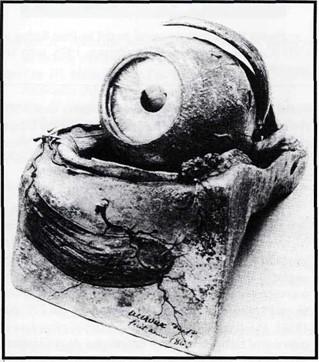 Figure 5. Anatomical model of the human eye by Auzoux, dated 1863. |
 Figure 6. Anatomical model by Auzoux, dated 1874 (Collection Librairie Alain Brieux, 1999, with kind permission). |
Anatomical models made of other materials
Bronze myologic statues date back to the Renaissance. Very few came down to our own day (Weil, no date). Some Italian names, chiefly Florentine artists, have been associated with these models: Francavilla, Marco d'Agrate and Lodovico Cigoli, among others.
| Whole body (man or woman), 130 movable parts, displaying 1700 structures | 3000 FF |
| Uterus with foetus | 500 FF |
| Woman pubis | 150 FF |
| Horse (1.20 m), 200 movable parts, displaying 300 structures | 2000 FF |
| June beetle, 12 x enlarged, displaying 600 structures | 250 FF |
Anatomical models made of sewn and padded material (silk) were used in Japan. Designations and some comments were painted (or embroidered?) on the different viscera (figure 7).
Paul Richer (1849-1933), professor of anatomy at the Ecole des Beaux-Arts, was famous for his researches in artistic anatomy. While assistant to Jean Martin Charcot at the Salpetriere, he published his first monograph on this subject (Richer, 1890). In the early twentieth century (ca. 1905), he produced plaster anatomical models of the human body (Brieux, 1996): a 61.5 cm standing man showing the superficial musculature on his left half (figure 8).
Since the early twentieth century, most of anatomical models were made of plastic or polychromatic rubber (figure 9).
 Figure 7. Anatomical model made of sewn and padded silk (taken from von Hagens and Whalley, 1995). |
 Figure 8. Plaster anatomical model by Paul Richer, ca. 1905 (Collection Librairie Alain Brieux, 1996, with kind permission). |
 Figure 9. Polychromatic rubber anatomical model of the human head, ca. 1935 (Collection Librairie Alain Brieux, 1995, with kind permission). |
Felice Fontana, Angelique Du Coudray, and Louis Auzoux had all the same purpose: they wanted to provide their students and colleagues with anatomical models that could be dismantled, or at least open, to display the most important structures of the human body. Moreover, these models also aimed at developing scientific popularization: " Only medical students had access to dissecting rooms. Thanks to him (Louis Auzoux), anatomy could be taught without the presence of death, in high schools and technical institutes. Thanks to him, paramedical students, especially midwives, could enjoy exceptionally realistic and bright instruction " (Saint-Restitut, 1986). Human dissection was acknowledged as being the best way to learn anatomy (Robert and Kiiss, 1840), but the pressing demand for human material exceeded by far the supply that was available (Olry, 1999). It was therefore necessary to make anatomical models available. Unfortunatelly, the price was often prohibitive: a model of a life-size standing man by Auzoux costed 3000 French francs, that is half of the annual salary of the Director of the Museum of that time! (Nowadays, a collector should pay out over 100,000 francs to buy the same model on the antiquarian market).
Though of high quality, there were inadequacies in all anatomical models. This problem was pointed out by Jean- Nicolas Gannal who wrote in 1838: " In brief, these three means to convey science [plates, wax models, and artificial anatomy] have their level of usefulness, but they could never bear comparison with the specific matter of organs; they could be useful to enrich a museum, but never to form it" (Gannal, 1838). Plastination has therefore to be regarded as the outcome of many centuries of educational determination: it combines qualities of anatomical models with authenticity of anatomical specimens.
Ameline JF: Me"moire sur l'utilite" des pieces d'anatomie artificielle chirurgicale. Paris: Fain, 1819.
Ameline JF: Memoire sur l'utilite des pieces d'anatomie artificielle chirurgicale. Caen: Le Roy, 1820.
Ameline JF: Notice sur de nouvelles pieces anatomiques artificielles, executees en carton, par M. Ameline, associe national a Caen. J gen Me"d Chir Pharm 81:28-31,1822.
Ameline JF: Observations sur les pieces d'anatomie d'Auzoux. Caen: Bonneserre, 1825.
Auzoux LTJ: [Reponse a M. Ameline]. Gazette des Hopitaux, February 9: 72,1839.
Brieux A: Histoire des sciences et de la me"decine. Livres anciens, documentation, instruments. Paris, November 1986.
Brieux A: Histoire des sciences et de la medecine. Livres anciens, documentation, instruments. Paris, December 1995.
Brieux A: Histoire des sciences et de la me"decine. Livres anciens, documentation, instruments, gravures & dessins. Paris, September 1996.
Brieux A: Histoire des sciences et de la medecine. Livres anciens, documentation, instruments. Paris, June 1999. Davis AB: Louis-Thomas-Jer6me Auzoux and the papier mache anatomical model. Riv Stor Sci med nat 20:257-279, 1977.
Fontana F: Courte description d'une statue anatomique qui se demonte en plus de 3000 parties. Mag Encycl 2: 35- 36, 1795.
Gannal JN: Histoire des embaumements et de la preparation des pieces d'anatomie normale, d'anatomie pathologique et d'histoire naturelle; suivie de precedes nouveaux. Paris: Ferra, 1838, pp. 194-198.
Grondin G, Olry R: Current Plastination Index. Trois-Rivieres: International Society for Plastination, 1996.
Hirsch A: Biographisches Lexicon der hervorragenden Aerzte aller Zeiten und Volker. Wien, Leipzig: Urban & Schwarzenberg, 1884-1888 (vol. 6 dated 1888).
Hugues S: Honore Fragonard l'anatomiste, les Ecorches au XVIIIe siecle. Paris, thesis. Jacquemin M: Compte-Rendu de l'anatomie clastique du Dr. Auzoux et de 1'influence qu'elle doit avoir sur l'instruction de la cavalerie. No place, no date.
Lauth T: Histoire de l'anatomie. Vol. 1 (all published). Strasbourg: F. G. Levrault, 1815.
Le Minor JM, Puygrenier J: La collection de cires anatomiques de l'Ecole du Service de Sante" des Armees de Lyon. Hist Sci Me"d 23 (2): 131-138, 1989.
Lemire M: Artistes et mortels. Paris: Raymond Chabeau, 1990
Olry R: Body Snatchers: the Hidden Side of the History of Anatomy. J Int Soc Plastination 14 (2): 6-9, 1999.
https://doi.org/10.56507/NXOD1218
Richer P: Traite d'anatomie artistique. Paris: Plon, 1890.
Robert A, Kiiss E: Anatomie humaine et comparee, moule"e en plStre sur nature et peintes d'apres les preparations. Strasbourg: G. Silbermann, 1840.
Saint-Restitut C: Le Dr Louis Auzoux. Un artisan du savoir. Gazette Med 93 (42): 71-73, 1986.
Saint-Restitut C: Angdlique Du Coudray, sage-femme itinerante, 1712-1789. Gazette Med 96 (18): 70-71,1989.
Sonolet J: A propos d'un mannequin anatomique en bois: Napoleon Bonaparte et Felice Fontana. Atti I. Congr.Internat. Ceroplastica. Florence, vol. 20, pp. 449-450, 1975.
https://doi.org/10.1017/S1446788700016165
Valsalva AM: De aure humana tractatus. Leiden: Apud Gisbertum Langerak & Johannem Hasebroek, 1735.
von Hagens G, Whalley A: Jintai no sekai [Human Body World]. 1995.
Wade RS: Medical Mummies: The History of the Burns Collection. Anat Rec: 158-161, 1998.
https://doi.org/10.1002/(SICI)1097-0185(199812)253:6<158::AID-AR3>3.0.CO;2-8
Weil E: Old Medicine. Catalogue 9. London, no date.